Mri scanner machine in radiology department
1344×768 · AVIF · CC BY 4.0
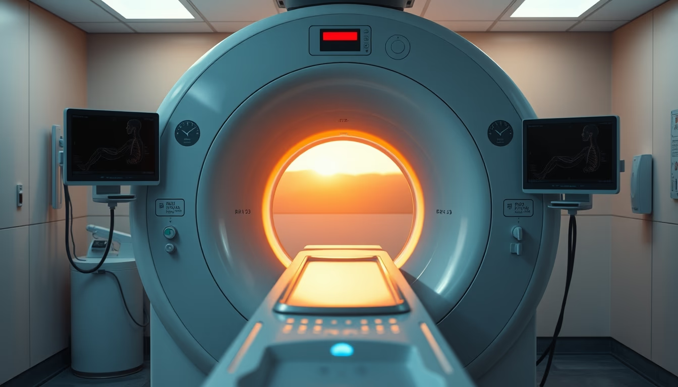
Magnetic resonance imaging is a non-invasive exam that uses magnetic fields and radio waves to produce detailed images of the human body.
About this subject
The magnetic resonance imaging (MRI) machine is one of the most advanced pieces of equipment in modern radiology. Unlike X-rays or computed tomography, MRI does not use ionizing radiation, making it safe for repeated exams, especially in children and pregnant women (although gadolinium contrast has restrictions). The physical principle is based on nuclear magnetic resonance: hydrogen protons in tissues are aligned by a strong magnetic field (usually 1.5 to 3 teslas) and excited by radiofrequency pulses; as they relax, they emit signals that are converted into images.
In the United States, MRI is regulated by the FDA and requires magnetically shielded rooms (Faraday cages) to prevent external interference. The first clinical MRI scanner was installed in the US in 1980 at the University of California, San Francisco. Since then, technology has evolved to include open MRI (reducing claustrophobia) and high-field systems (7T) for research.
The exam is indicated for diagnosing neurological disorders (such as multiple sclerosis and tumors), orthopedic injuries (ligament and meniscus tears), cardiac conditions, and abdominal diseases. The duration ranges from 20 to 60 minutes, depending on the area. Patients must remain still to avoid motion artifacts. Loud noises (up to 120 dB) require ear protection.
A curiosity: liquid helium is used to cool superconducting magnets to near absolute zero (-269°C). Without this system, the magnetic field would not remain stable. Additionally, functional MRI (fMRI) can map brain activity in real time, making it an essential tool in neuroscience.
Frequently Asked Questions
What is the difference between MRI and CT scan?
MRI uses magnetic fields and radio waves without ionizing radiation, while CT uses X-rays. MRI provides better soft tissue contrast but is slower and more sensitive to motion.
Who cannot have an MRI?
Patients with pacemakers, ferromagnetic metal implants (e.g., old aneurysm clips), or intraocular metal fragments cannot undergo MRI. MRI-conditional devices are allowed under specific protocols.
Does an MRI hurt?
No, the exam is painless. Discomfort may arise from lying still for a long time or claustrophobia inside the bore. Mild sedation is sometimes used for anxious patients.
Direct URL
https://pub-c7d6a6ea828543ac903a74a341ccb2e1.r2.dev/imagens/mri-scanner-machine-in-radiology-department-symmetric-composition-golden-hour-late-afternoon.avifHow to credit
Include a visible link back to UtilizAí. Copy one of the snippets below:
<a href="https://xn--utiliza-eza.com/en/midia/imagens/mri-scanner-machine-in-radiology-department-symmetric-composition-golden-hour-late-afternoon">Mri scanner machine in radiology department</a> by <a href="https://xn--utiliza-eza.com">UtilizAí</a>, licensed under <a href="https://creativecommons.org/licenses/by/4.0/">CC BY 4.0</a>.
[Mri scanner machine in radiology department](https://xn--utiliza-eza.com/en/midia/imagens/mri-scanner-machine-in-radiology-department-symmetric-composition-golden-hour-late-afternoon) by [UtilizAí](https://xn--utiliza-eza.com), CC BY 4.0
License: CC-BY-4.0