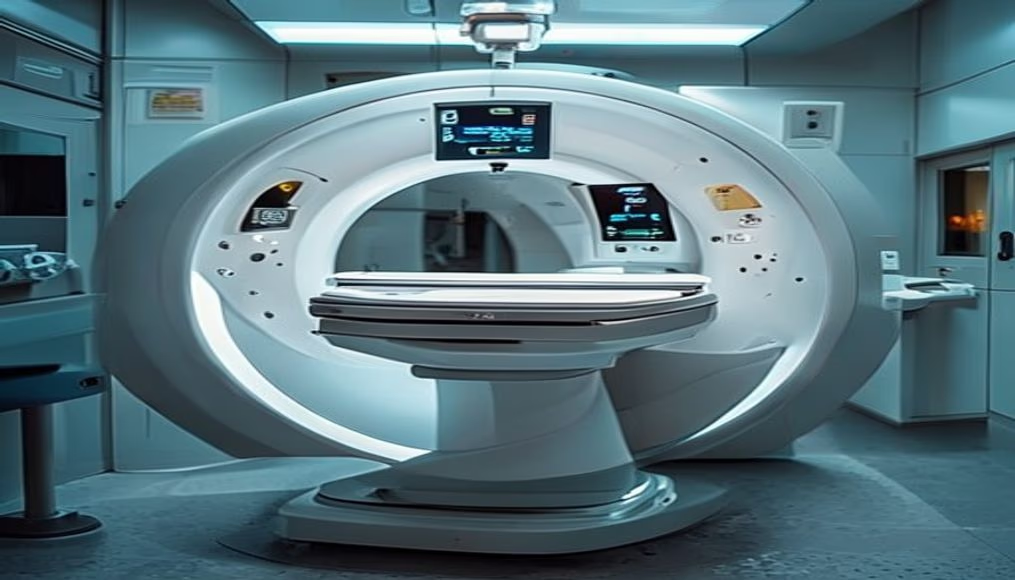
Mri scanner machine in radiology department
1344×768 · AVIF · CC BY 4.0
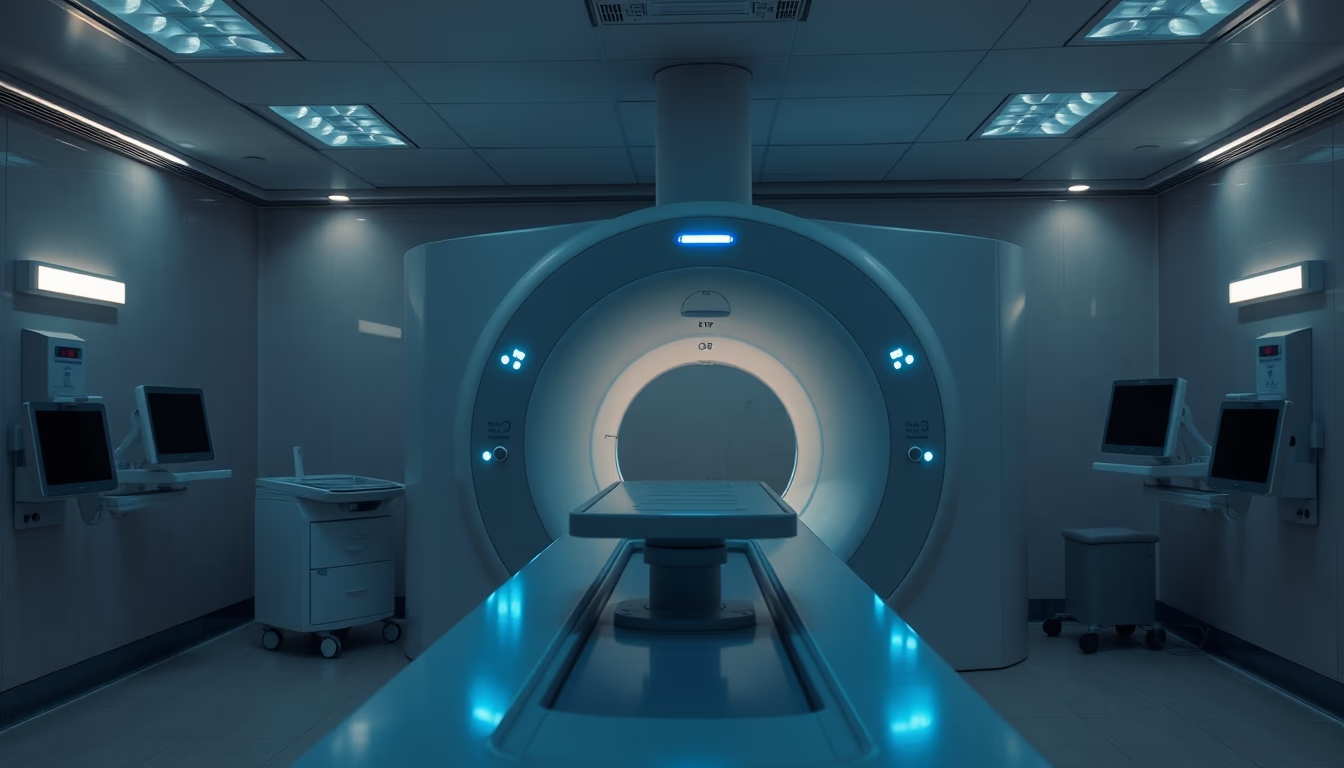
An MRI scanner machine in a radiology department, highlighting diagnostic imaging technology in a hospital setting.
About this subject
Magnetic resonance imaging (MRI) is a non-invasive medical imaging technique that uses magnetic fields and radio waves to generate detailed images of organs and tissues. Unlike computed tomography (CT), MRI does not use ionizing radiation, making it particularly useful for visualizing soft tissues such as the brain, spinal cord, muscles, and joints. The equipment consists of a large tunnel-shaped magnet where the patient is positioned for the scan. The first MRI scanners were developed in the 1970s, with the first human scan performed in 1977. Since then, technology has evolved significantly, with stronger magnetic fields (1.5T, 3T, and even 7T in research) and faster pulse sequences, reducing scan time and improving image quality.
In Brazil, MRI is widely available in public and private hospitals, regulated by the National Health Surveillance Agency (ANVISA). The exam is indicated for diagnosing various conditions, such as tumors, neurological diseases (multiple sclerosis, Alzheimer's), musculoskeletal injuries, and cardiovascular problems. Safety is a major concern: patients with metal implants, pacemakers, or aneurysm clips may have contraindications due to the strong magnetic field. Therefore, rigorous screening is performed before the procedure.
The image, with blue lighting and a wide composition, evokes the nighttime environment of a hospital, where emergency exams are performed. The twilight blue hour tone gives a serene and technological atmosphere, contrasting with the complexity of the equipment. MRI is a symbol of modern medicine, representing advances in early and non-invasive diagnosis.
Frequently Asked Questions
Is MRI safe? Are there any risks?
MRI is considered safe as it does not use ionizing radiation. However, the strong magnetic field can be dangerous for people with metal implants, pacemakers, or aneurysm clips. It is essential to inform the doctor about any devices before the exam.
How long does an MRI scan take?
The duration ranges from 20 to 60 minutes, depending on the body part and the type of sequences used. More complex exams, such as cardiac MRI, can take up to 90 minutes.
What is the difference between MRI and CT scan?
MRI uses magnetic fields and radio waves, being better for soft tissues. CT uses X-rays, being faster and indicated for bones and emergencies. MRI does not emit radiation, while CT exposes the patient to a dose of radiation.
Direct URL
https://pub-c7d6a6ea828543ac903a74a341ccb2e1.r2.dev/imagens/mri-scanner-machine-in-radiology-department-cinematic-wide-shot-twilight-blue-hour.avifHow to credit
Include a visible link back to UtilizAí. Copy one of the snippets below:
<a href="https://xn--utiliza-eza.com/en/midia/imagens/mri-scanner-machine-in-radiology-department-cinematic-wide-shot-twilight-blue-hour">Mri scanner machine in radiology department</a> by <a href="https://xn--utiliza-eza.com">UtilizAí</a>, licensed under <a href="https://creativecommons.org/licenses/by/4.0/">CC BY 4.0</a>.
[Mri scanner machine in radiology department](https://xn--utiliza-eza.com/en/midia/imagens/mri-scanner-machine-in-radiology-department-cinematic-wide-shot-twilight-blue-hour) by [UtilizAí](https://xn--utiliza-eza.com), CC BY 4.0
License: CC-BY-4.0