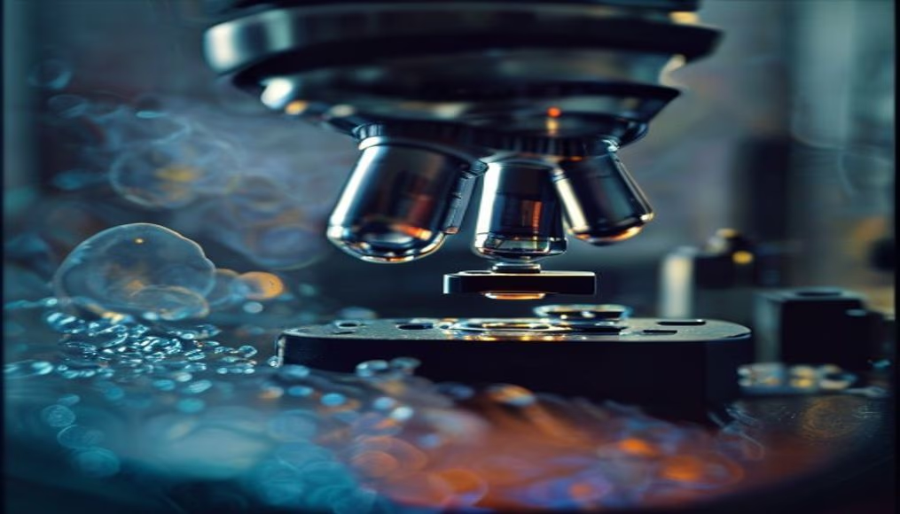
Microscope view cell culture
1344×768 · AVIF · CC BY 4.0
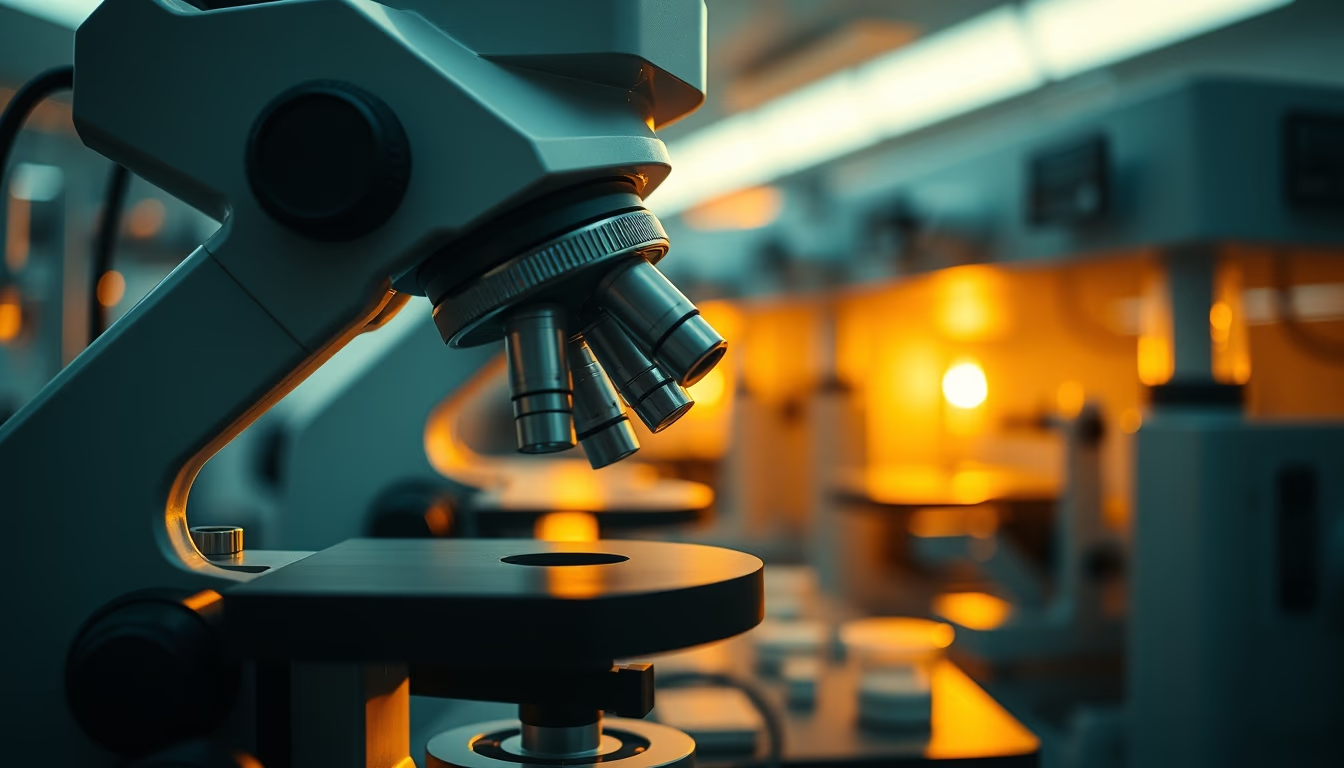
Microscopy of cell cultures is essential for research in biology, medicine, and pharmacology, enabling real-time observation of cellular behavior.
About this subject
Cell culture is a fundamental technique in laboratories worldwide, used to study biological processes, test drugs, and produce vaccines. Microscopic visualization allows scientists to monitor cell growth, division, and response to various stimuli. This practice is common in fields such as oncology, where the effect of drugs on tumor cells is observed, and virology, to track cellular infection.
Various types of microscopy are applied to cell cultures, such as phase-contrast microscopy, which enhances details without staining, and fluorescence microscopy, which uses markers to identify specific proteins. Technology has advanced to include time-lapse systems that capture images at regular intervals, revealing cellular dynamics that would be imperceptible in single observations.
In Brazil, research centers like the Butantan Institute and Fiocruz heavily use cell cultures for developing sera and vaccines. The technique is also crucial for producing monoclonal antibodies and cell therapies. Maintaining sterile conditions and controlling temperature and CO2 levels are vital to ensure cell viability, which can be from immortalized lines or primary cells extracted directly from tissues.
Interestingly, the first successful cell culture was performed in 1907 by American embryologist Ross Harrison, who cultured frog nerve cells. Since then, the technique has evolved to include synthetic culture media and bioreactors that enable large-scale production. The image of a microscope over a cell culture represents the link between the microscopic world and life-saving discoveries.
Frequently Asked Questions
What is cell culture and what is it used for?
Cell culture is the process of growing cells in a controlled environment outside the organism. It is used for biological research, drug testing, vaccine production, and cell therapies.
What types of microscopy are used in cell cultures?
The main types are phase-contrast microscopy, which observes living cells without staining, and fluorescence microscopy, which uses dyes to mark specific structures.
How is a cell culture maintained?
Cells are kept in culture media with nutrients, in incubators with controlled temperature (37°C) and CO2 (5%), in a sterile environment to prevent contamination.
Direct URL
https://pub-c7d6a6ea828543ac903a74a341ccb2e1.r2.dev/imagens/microscope-view-cell-culture-cinematic-wide-shot-v2.avifHow to credit
Include a visible link back to UtilizAí. Copy one of the snippets below:
<a href="https://xn--utiliza-eza.com/en/midia/imagens/microscope-view-cell-culture-cinematic-wide-shot-v2">Microscope view cell culture</a> by <a href="https://xn--utiliza-eza.com">UtilizAí</a>, licensed under <a href="https://creativecommons.org/licenses/by/4.0/">CC BY 4.0</a>.
[Microscope view cell culture](https://xn--utiliza-eza.com/en/midia/imagens/microscope-view-cell-culture-cinematic-wide-shot-v2) by [UtilizAí](https://xn--utiliza-eza.com), CC BY 4.0
License: CC-BY-4.0