Electron microscope sample
1344×768 · AVIF · CC BY 4.0
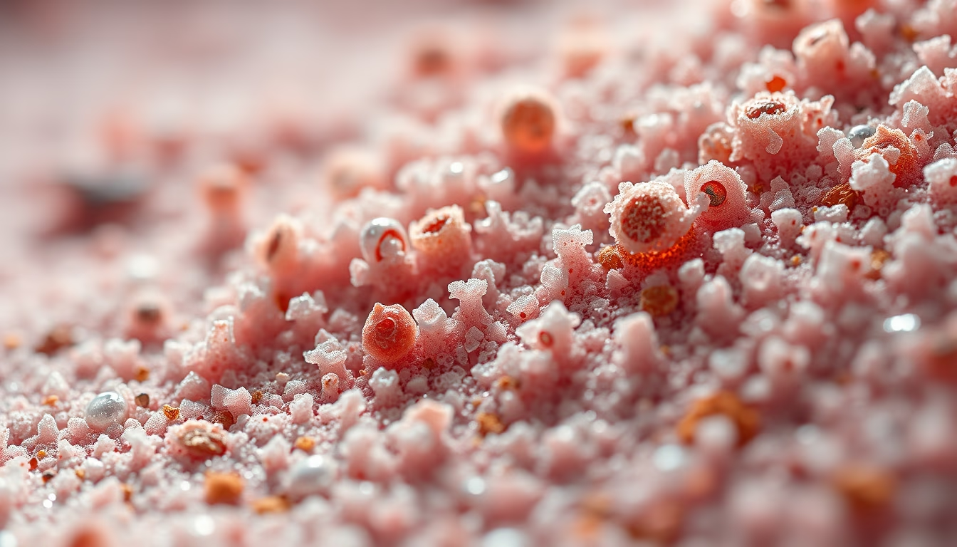
Samples observed under electron microscopes reveal structures invisible to the naked eye, combining science and art in ultra-high-resolution images.
About this subject
Electron microscopes use beams of electrons instead of light to magnify objects, achieving resolutions as fine as 0.1 nanometer, far beyond conventional optical microscopes. Biological samples, such as cells, tissues, or microorganisms, must undergo a rigorous preparation process: chemical fixation, dehydration, resin embedding, and ultra-thin sectioning (for transmission microscopy) or metal coating with gold or platinum (for scanning microscopy). This preparation is essential to withstand the vacuum and electron bombardment while preserving the original morphology.
Electron microscopy revolutionized biology by enabling the visualization of cellular organelles, viruses, and macromolecules. Discoveries such as the structure of the ribosome, the organization of the cytoskeleton, and the morphology of pathogens like SARS-CoV-2 were only possible thanks to this technique. Beyond scientific value, the resulting images, with their textures, shadows, and metallic contrasts, are often exhibited as photographic art, highlighting the hidden beauty of nature at microscopic scales.
In the realm of fine-art photography, these micrographs transcend scientific documentation, appreciated for their abstract aesthetics and detailed composition. Artists and researchers collaborate to enhance false colors and angles, creating works that spark curiosity about the invisible world. The combination of technical precision and artistic expression makes each sample a unique piece, bridging laboratories and art galleries.
Interestingly, even non-biological samples, such as crystals or materials, gain new life under the electron microscope, revealing surprising geometric patterns. This intersection of science and art underscores the importance of cutting-edge equipment and human creativity in exploring the infinitely small.
Frequently Asked Questions
How are samples prepared for electron microscopy?
Samples undergo chemical fixation, dehydration, resin embedding, and ultra-thin sectioning (for TEM) or metal coating with gold (for SEM) to withstand vacuum and electron beams.
What is the difference between SEM and TEM?
SEM scans the sample surface with electrons, producing 3D topographical images. TEM transmits electrons through the sample, revealing internal structures in thin sections.
Why are electron microscopy images considered art?
Because they combine scientific precision with visual composition, metallic textures, and contrasts reminiscent of abstractions. Many are artificially colored and displayed in galleries as fine art.
Direct URL
https://pub-c7d6a6ea828543ac903a74a341ccb2e1.r2.dev/imagens/electron-microscope-sample-fine-art-photography-p7.avifHow to credit
Include a visible link back to UtilizAí. Copy one of the snippets below:
<a href="https://xn--utiliza-eza.com/en/midia/imagens/electron-microscope-sample-fine-art-photography-p7">Electron microscope sample</a> by <a href="https://xn--utiliza-eza.com">UtilizAí</a>, licensed under <a href="https://creativecommons.org/licenses/by/4.0/">CC BY 4.0</a>.
[Electron microscope sample](https://xn--utiliza-eza.com/en/midia/imagens/electron-microscope-sample-fine-art-photography-p7) by [UtilizAí](https://xn--utiliza-eza.com), CC BY 4.0
License: CC-BY-4.0





