Electron microscope sample
1344×768 · AVIF · CC BY 4.0
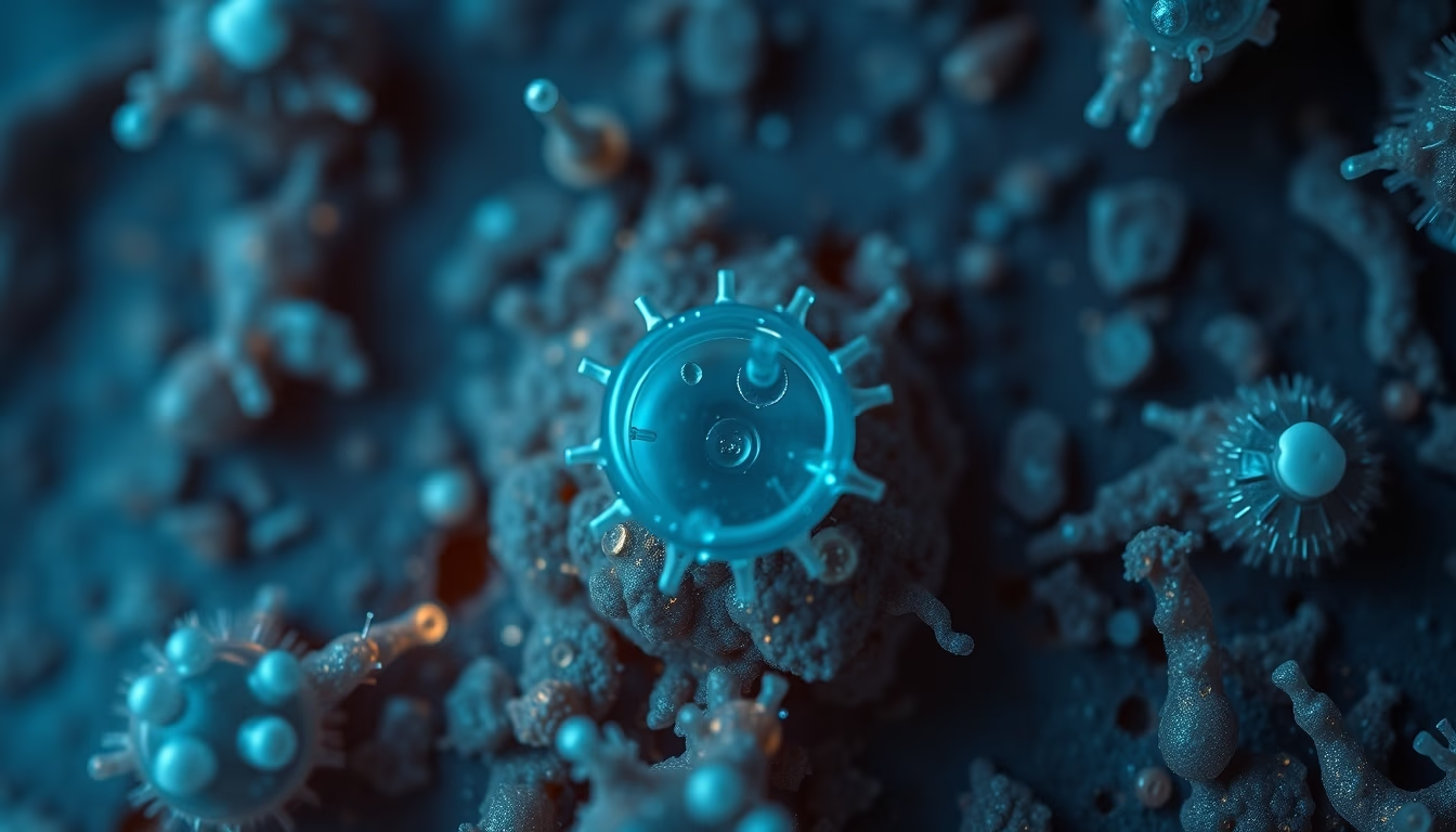
Samples for electron microscopy require meticulous preparation, revealing structures at the nanoscale.
About this subject
Electron microscopy uses a beam of electrons to image samples with resolution far surpassing light microscopy, achieving magnifications up to 10 million times. Samples must be ultrathin (below 100 nanometers) and conductive, often coated with gold or carbon. Preparation involves chemical fixation, dehydration, resin embedding, and sectioning with an ultramicrotome. For biological samples, cryofixation preserves native structures. Scanning electron microscopy (SEM) produces three-dimensional surface images, while transmission electron microscopy (TEM) reveals internal details. Electron energy loss spectroscopy (EELS) coupled with TEM enables chemical analysis at the atomic scale. Challenges include radiation damage and preparation artifacts, requiring researcher expertise. Advances in detectors and software improve image acquisition and processing. Applications range from materials science to cell biology, allowing visualization of viruses, proteins, and crystal defects.
Frequently Asked Questions
What is the difference between scanning electron microscopy (SEM) and transmission electron microscopy (TEM)?
SEM produces surface images with a 3D appearance, while TEM reveals internal structures by transmitting electrons through an ultrathin sample.
Why must samples be conductive for electron microscopy?
Incident electrons can cause charge buildup on the sample, distorting the image. Conductive coatings like gold or carbon dissipate this charge.
What is the ideal thickness for a transmission electron microscopy sample?
Typically between 50 and 100 nanometers, allowing electrons to pass through and form a clear image.
Direct URL
https://pub-c7d6a6ea828543ac903a74a341ccb2e1.r2.dev/imagens/electron-microscope-sample-aerial-drone-shot-p4.avifHow to credit
Include a visible link back to UtilizAí. Copy one of the snippets below:
<a href="https://xn--utiliza-eza.com/en/midia/imagens/electron-microscope-sample-aerial-drone-shot-p4">Electron microscope sample</a> by <a href="https://xn--utiliza-eza.com">UtilizAí</a>, licensed under <a href="https://creativecommons.org/licenses/by/4.0/">CC BY 4.0</a>.
[Electron microscope sample](https://xn--utiliza-eza.com/en/midia/imagens/electron-microscope-sample-aerial-drone-shot-p4) by [UtilizAí](https://xn--utiliza-eza.com), CC BY 4.0
License: CC-BY-4.0





