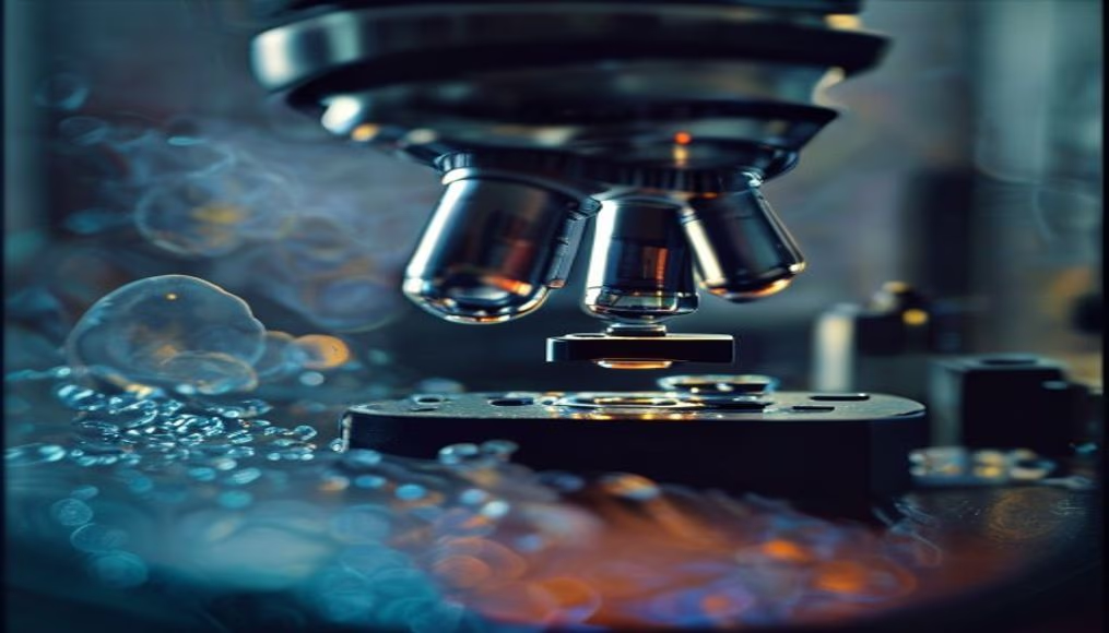
Cells under microscope
1344×768 · AVIF · CC BY 4.0

Cells are the fundamental units of life, and observing them under a microscope reveals complex structures essential for medical diagnostics and scientific research.
About this subject
Cells are the structural and functional basis of all living organisms. They are broadly classified into prokaryotic cells, which lack a defined nucleus (e.g., bacteria), and eukaryotic cells, which have an organized nucleus (found in animals, plants, and fungi). The average size of a eukaryotic cell ranges from 10 to 100 micrometers, invisible to the naked eye, making the microscope an essential tool for their study.
The history of microscopy began in the 17th century with Antonie van Leeuwenhoek, who observed protozoa and bacteria using simple lenses. Later advances, such as the compound microscope and the electron microscope, enabled visualization of organelles like mitochondria and ribosomes. The cell theory, formulated in the 19th century, established that all living things are composed of cells and that cells arise only from pre-existing cells, revolutionizing biology.
In healthcare, microscopic observation of cells is critical for diagnosis. Blood tests, biopsies, and Pap smears rely on cellular morphology to identify diseases such as anemia, infections, and cancer. Cancer cells, for instance, exhibit enlarged nuclei and irregular shapes, features that pathologists recognize to determine tumor type and grade. Fluorescence microscopy further allows labeling of specific proteins, aiding in drug research and development.
Interestingly, an adult human body contains about 37 trillion cells, each with specialized functions. Stem cells, capable of differentiating into various tissues, are studied for organ regeneration. Cell division through mitosis or meiosis ensures growth and reproduction. Even after death, some skin and hair cells continue dividing for hours, a phenomenon exploited in transplants.
Frequently Asked Questions
What are cells and why are they important?
Cells are the smallest functional units of living organisms. They perform all vital functions such as metabolism, reproduction, and response to stimuli, forming the basis of life.
How does a microscope allow us to see cells?
Optical microscopes use lenses to magnify the image, while electron microscopes use electron beams. To observe details, cells are often stained with specific dyes that contrast their structures.
Why is the study of cells important in medicine?
Cell study enables diagnosis of diseases like cancer and infections, understanding of drug mechanisms, and development of advanced therapies such as cell therapy and gene editing.
Direct URL
https://pub-c7d6a6ea828543ac903a74a341ccb2e1.r2.dev/imagens/cells-under-microscope-travel-photography-v10.avifHow to credit
Include a visible link back to UtilizAí. Copy one of the snippets below:
<a href="https://xn--utiliza-eza.com/en/midia/imagens/cells-under-microscope-travel-photography-v10">Cells under microscope</a> by <a href="https://xn--utiliza-eza.com">UtilizAí</a>, licensed under <a href="https://creativecommons.org/licenses/by/4.0/">CC BY 4.0</a>.
[Cells under microscope](https://xn--utiliza-eza.com/en/midia/imagens/cells-under-microscope-travel-photography-v10) by [UtilizAí](https://xn--utiliza-eza.com), CC BY 4.0
License: CC-BY-4.0