Cells under microscope
1344×768 · AVIF · CC BY 4.0
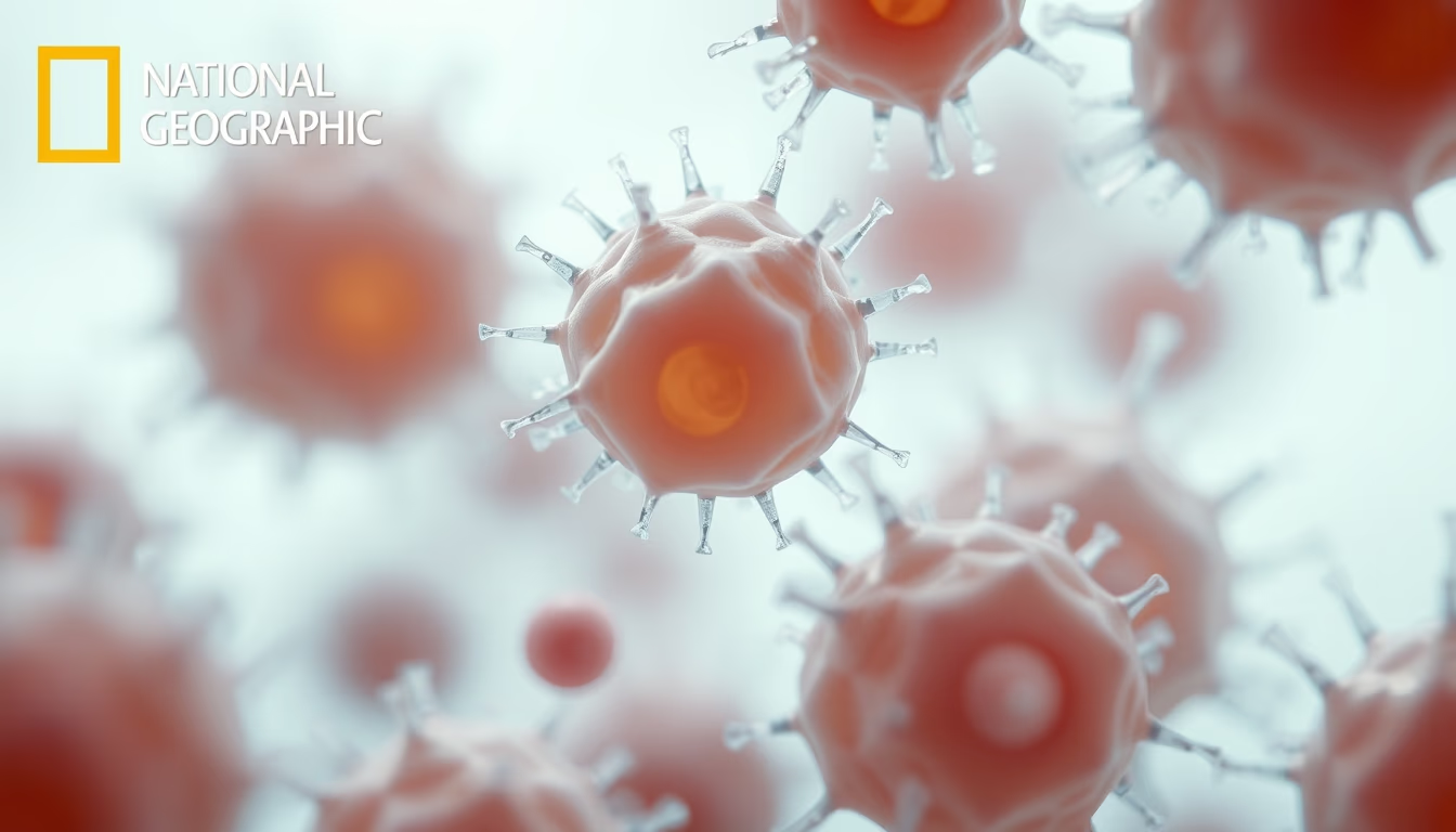
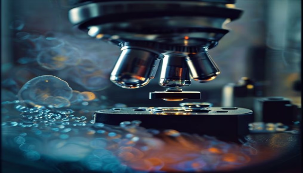
Human cells viewed under a microscope reveal complex structures and vital processes that sustain life.
About this subject
Cells are the fundamental units of life, and their observation under a microscope revolutionized biology starting in the 17th century, when Robert Hooke first described cork cells. Today, fluorescence and electron microscopes allow visualization of organelles such as the nucleus, mitochondria, and endoplasmic reticulum in nanometer detail. Each human cell contains about 20,000 to 25,000 genes in the nuclear DNA, which coordinate the production of essential proteins. Mitochondria, known as powerhouses, convert glucose into ATP through cellular respiration, a process that generates approximately 90% of the body's energy. Cell study is crucial for understanding diseases like cancer, where genetic mutations lead to uncontrolled proliferation. Techniques such as confocal microscopy enable three-dimensional reconstructions of tissues, aiding early diagnosis. Furthermore, the discovery of induced pluripotent stem cells (iPS) in 2006 by Shinya Yamanaka opened avenues for regenerative medicine. The image of cells under a microscope is not just a visual record but a window into the molecular complexity that defines our existence.
Frequently Asked Questions
What are the main organelles visible under a light microscope?
Under a standard light microscope, you can see the nucleus (stained), vacuoles, and sometimes mitochondria and chloroplasts (in plant cells). Smaller organelles like ribosomes require electron microscopy.
How do cells communicate with each other?
Cells communicate through chemical signals such as hormones and neurotransmitters that bind to membrane receptors. Direct contact also occurs via gap junctions in animal tissues.
What is the difference between prokaryotic and eukaryotic cells?
Prokaryotic cells (bacteria) lack a defined nucleus and membrane-bound organelles. Eukaryotic cells (animals, plants, fungi) have a nucleus enclosed by a membrane and organelles like mitochondria and endoplasmic reticulum.
Direct URL
https://pub-c7d6a6ea828543ac903a74a341ccb2e1.r2.dev/imagens/cells-under-microscope-national-geographic-style.avifHow to credit
Include a visible link back to UtilizAí. Copy one of the snippets below:
<a href="https://xn--utiliza-eza.com/en/midia/imagens/cells-under-microscope-national-geographic-style">Cells under microscope</a> by <a href="https://xn--utiliza-eza.com">UtilizAí</a>, licensed under <a href="https://creativecommons.org/licenses/by/4.0/">CC BY 4.0</a>.
[Cells under microscope](https://xn--utiliza-eza.com/en/midia/imagens/cells-under-microscope-national-geographic-style) by [UtilizAí](https://xn--utiliza-eza.com), CC BY 4.0
License: CC-BY-4.0