Cell division microscope
1344×768 · AVIF · CC BY 4.0
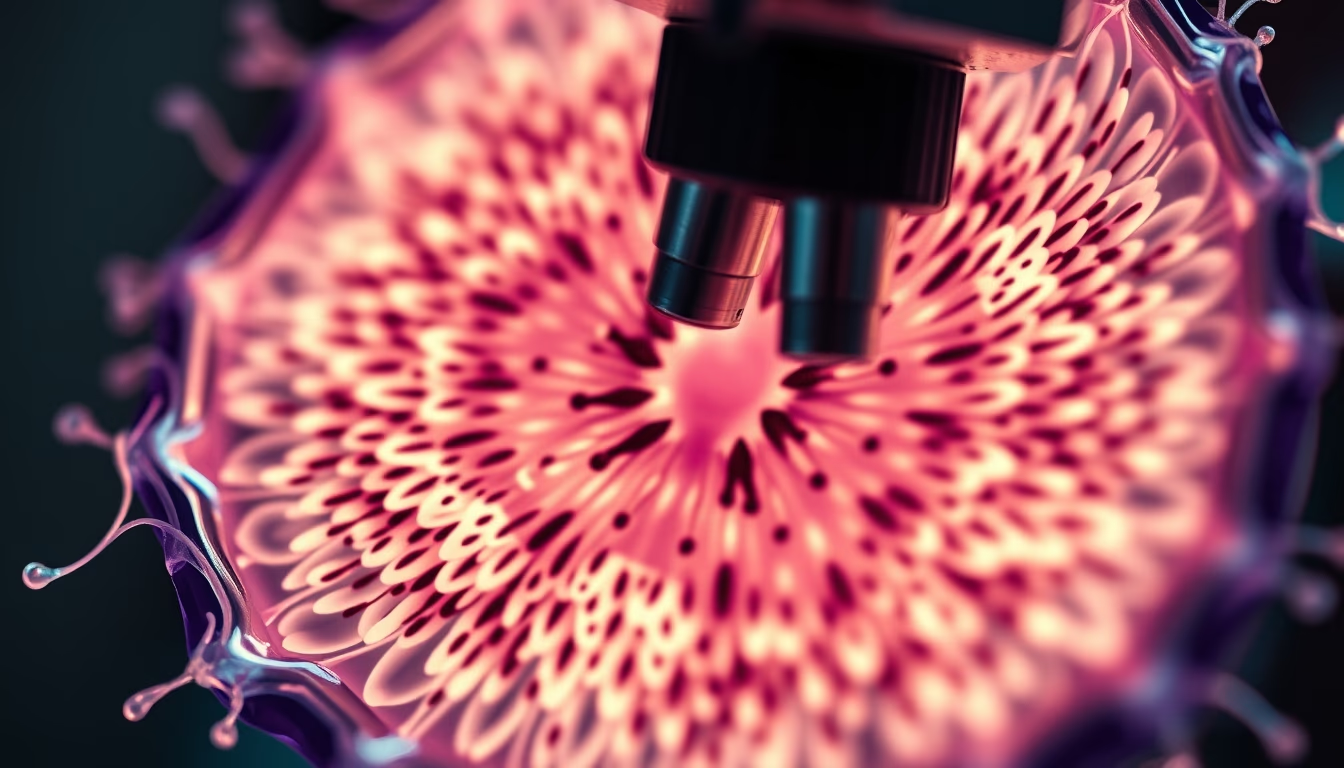
Cell division, observed through microscopy, is the fundamental process by which a cell replicates itself, essential for tissue growth and repair.
About this subject
Cell division is an essential biological process that occurs in all living organisms. In eukaryotes, mitosis is the most common type, responsible for producing two daughter cells genetically identical to the parent cell. This process is crucial for growth, repair of damaged cells, and asexual reproduction. Analysis of cell division by optical and electron microscopy has led to significant advances in understanding the cell cycle, with emphasis on the phases: prophase, metaphase, anaphase, and telophase. Errors in cell division, such as chromosomal nondisjunction, can lead to aneuploidies, often associated with conditions like Down syndrome and various types of cancer.
Fluorescence microscopy revolutionized the study of cell division, enabling visualization of specific proteins involved in spindle formation and chromosome segregation. Scientists like Theodor Boveri and Walter Sutton, in the early 20th century, established the chromosomal basis of heredity through detailed observations of meiosis. Today, high-resolution microscopy and time-lapse techniques allow tracking the process in living cells, revealing previously invisible molecular dynamics.
In addition to mitosis, meiosis is another type of cell division occurring in germ cells, reducing chromosome number by half to form gametes. Detailed understanding of cell division has direct implications for developing anticancer therapies, which often target cell cycle regulatory proteins. Artistic imaging of cell division through microscopy combines science and aesthetics, showcasing the intrinsic beauty of fundamental biological processes.
Frequently Asked Questions
What are the main phases of mitosis?
Mitosis is divided into prophase, metaphase, anaphase, and telophase. In prophase, chromosomes condense and the mitotic spindle begins to form. In metaphase, chromosomes align at the cell equator. In anaphase, sister chromatids separate and are pulled to opposite poles. In telophase, nuclear membranes reform around the two sets of chromosomes.
What is the difference between mitosis and meiosis?
Mitosis produces two daughter cells genetically identical to the parent cell (diploid), while meiosis produces four daughter cells with half the chromosome number (haploid). Meiosis involves two consecutive divisions and is specific for gamete formation, essential for sexual reproduction.
How does microscopy contribute to the study of cell division?
Microscopy allows direct visualization of cellular structures during division. Techniques like fluorescence and phase-contrast microscopy enable observation of chromosomes, mitotic spindle, and protein dynamics in real time, crucial for understanding molecular mechanisms and detecting abnormalities associated with diseases.
Direct URL
https://pub-c7d6a6ea828543ac903a74a341ccb2e1.r2.dev/imagens/cell-division-microscope-fine-art-photography-p11.avifHow to credit
Include a visible link back to UtilizAí. Copy one of the snippets below:
<a href="https://xn--utiliza-eza.com/en/midia/imagens/cell-division-microscope-fine-art-photography-p11">Cell division microscope</a> by <a href="https://xn--utiliza-eza.com">UtilizAí</a>, licensed under <a href="https://creativecommons.org/licenses/by/4.0/">CC BY 4.0</a>.
[Cell division microscope](https://xn--utiliza-eza.com/en/midia/imagens/cell-division-microscope-fine-art-photography-p11) by [UtilizAí](https://xn--utiliza-eza.com), CC BY 4.0
License: CC-BY-4.0





