Bacterial colony petri dish
1344×768 · AVIF · CC BY 4.0
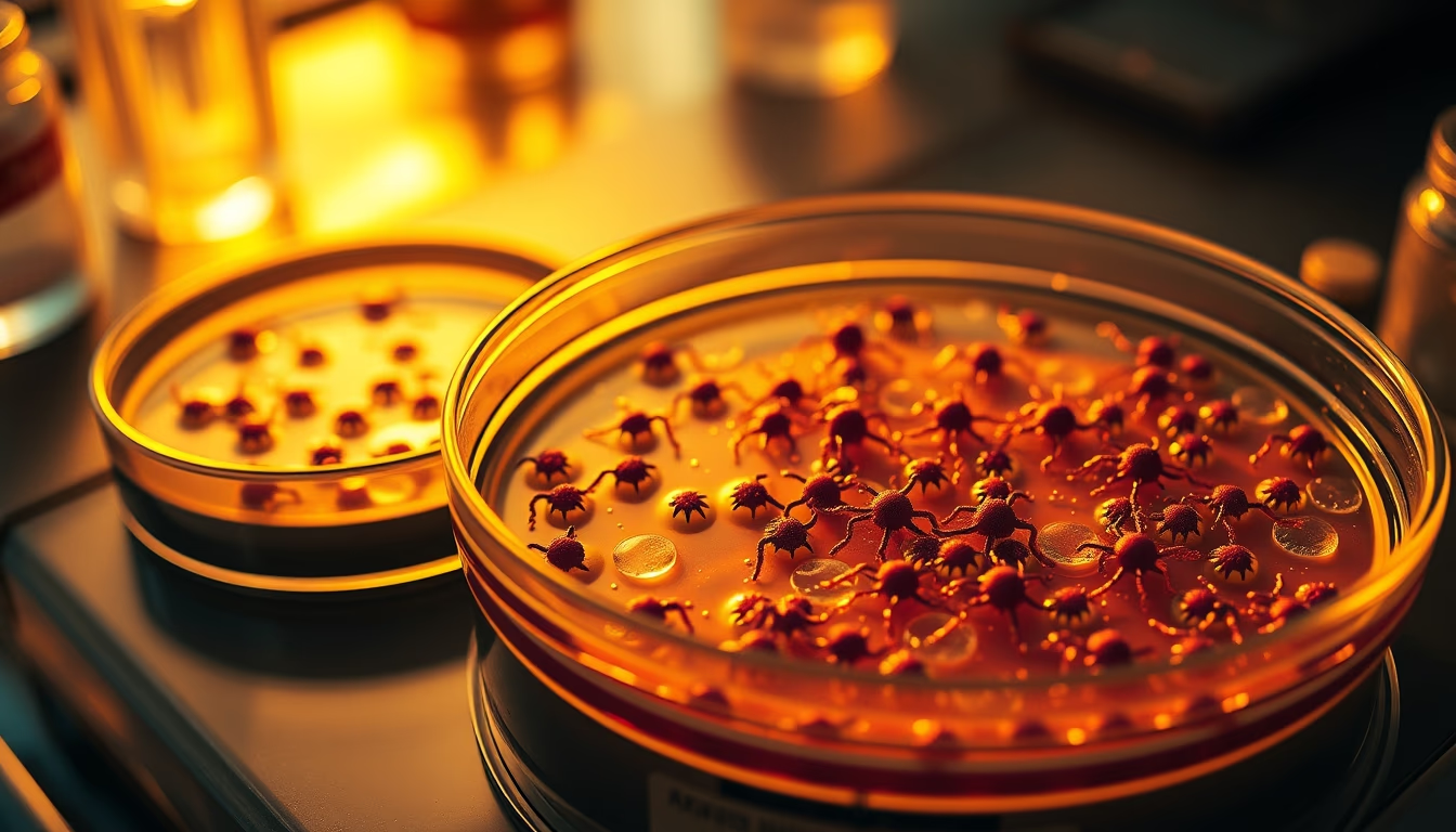
A petri dish with bacterial colonies reveals microbial diversity, essential for clinical and industrial microbiology studies.
About this subject
The Petri dish, invented by German bacteriologist Julius Richard Petri in 1887, is one of the most iconic tools in microbiology. It consists of a circular glass or plastic container with a lid, filled with a solid culture medium such as nutrient agar. Bacteria are inoculated onto the medium and, after incubation, form visible colonies. Each colony is a clone of millions of cells derived from a single original cell, exhibiting morphological characteristics like color, shape, edge, and texture that aid in preliminary identification of microorganisms.
Bacterial colonies vary widely: they can be round, irregular, mucoid, dry, pigmented, or translucent. For example, Serratia marcescens produces a red pigment called prodigiosin, while Pseudomonas aeruginosa secretes pyocyanin, a blue-green color. These features are used in clinical labs to differentiate common pathogens such as Staphylococcus aureus (golden colonies) and Escherichia coli (pink colonies on MacConkey agar). Colony morphology is a first step in bacterial identification, later confirmed by biochemical or molecular tests.
Petri dishes are essential beyond identification. They are key in antimicrobial susceptibility testing, such as the disk diffusion method (Kirby-Bauer), where antibiotic disks are placed on agar to measure inhibition zones. In the food industry, plates are used for counting indicator microorganisms like coliforms. In academia, culturing on plates allows isolation of microbes from soil, water, or air, revealing immense microbial biodiversity. It is estimated that only 1% of soil bacteria are culturable in the lab, highlighting the discovery potential with improved techniques.
Interestingly, the inoculation process requires strict aseptic technique to avoid contamination. Streaking for isolation, for example, using the quadrant method, yields single colonies from a mixed sample. Each colony represents a colony-forming unit (CFU), used to quantify viable bacteria. Careful observation of colony characteristics, size, elevation, margin, and sheen, is a skill microbiologists develop over years, making the Petri dish a simple yet powerful tool to understand the invisible world around us.
Frequently Asked Questions
What is a bacterial colony?
A bacterial colony is a visible cluster of millions of genetically identical bacteria, originating from a single cell that has repeatedly divided on a solid culture medium.
How are bacterial colonies formed in a petri dish?
After inoculating a sample containing bacteria onto the agar and incubating under suitable conditions, each bacterial cell multiplies to form an isolated colony. Colony morphology depends on the species and growth conditions.
What are petri dishes used for in microbiology?
They are used to isolate, culture, and identify microorganisms, test antibiotic sensitivity, quantify bacteria in samples (such as food or water), and study metabolic and morphological characteristics.
Direct URL
https://pub-c7d6a6ea828543ac903a74a341ccb2e1.r2.dev/imagens/bacterial-colony-petri-dish-cinematic-wide-shot-p2.avifHow to credit
Include a visible link back to UtilizAí. Copy one of the snippets below:
<a href="https://xn--utiliza-eza.com/en/midia/imagens/bacterial-colony-petri-dish-cinematic-wide-shot-p2">Bacterial colony petri dish</a> by <a href="https://xn--utiliza-eza.com">UtilizAí</a>, licensed under <a href="https://creativecommons.org/licenses/by/4.0/">CC BY 4.0</a>.
[Bacterial colony petri dish](https://xn--utiliza-eza.com/en/midia/imagens/bacterial-colony-petri-dish-cinematic-wide-shot-p2) by [UtilizAí](https://xn--utiliza-eza.com), CC BY 4.0
License: CC-BY-4.0





