Bacterial colony petri dish
1344×768 · AVIF · CC BY 4.0
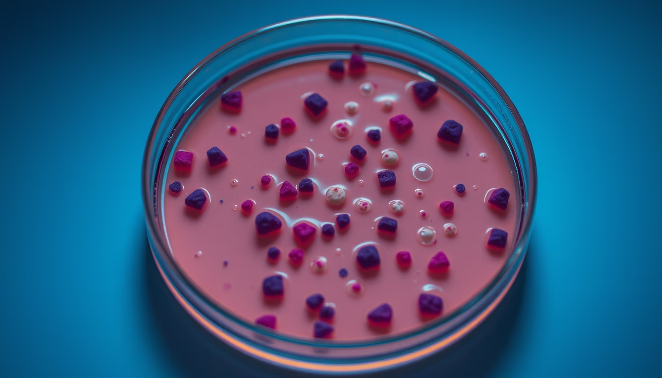
Bacterial colonies on a Petri dish reveal microbial growth patterns essential for laboratory studies and clinical diagnostics.
About this subject
Bacterial colonies are visible clusters of microorganisms originating from a single cell, grown on solid media such as agar inside Petri dishes. This method, developed by German bacteriologist Julius Petri in 1887, revolutionized microbiology by allowing the isolation and observation of pure species. The process begins with inoculating a sample onto the agar surface using techniques like streak plating, which progressively dilutes the microorganisms until individual cells form separate colonies.
Colony morphology, size, shape, color, margin, elevation, and texture, provides valuable clues for preliminary bacterial identification. For instance, mucoid colonies indicate capsule production, while irregular margins may suggest motility. Additionally, selective and differential media, such as MacConkey agar, inhibit certain groups and reveal metabolic characteristics, like lactose fermentation that alters colony color.
In clinical practice, Petri dish culture is essential for diagnosing bacterial infections, testing antibiotic susceptibility (antibiogram), and monitoring contamination in food and hospital environments. The technique is also widely used in research to study bacterial genetics, the action of new drugs, and microbial ecology. Interestingly, colony morphology can be so specific that experienced microbiologists often identify common genera just by colony appearance, even before biochemical tests.
Frequently Asked Questions
How do you streak a Petri dish to obtain isolated colonies?
Use a sterile loop, touch the sample, and streak in a zigzag pattern over one quadrant. Flame the loop, rotate the plate 90 degrees, and streak from the previous quadrant into the next, repeating for four quadrants. This dilutes the sample, yielding isolated colonies in the last quadrant.
Why do bacterial colonies have different colors?
Colors can come from natural pigments produced by the bacterium (e.g., Serratia marcescens produces red) or from indicators in the culture medium, such as phenol red that changes color with sugar fermentation. Color aids in presumptive identification.
What is the importance of colony morphology in bacterial identification?
Colony morphology (size, shape, margin, elevation, texture, color) is one of the first steps in bacterial identification. It guides the choice of biochemical tests and can differentiate common genera, such as Staphylococcus (opaque, smooth colonies) and Streptococcus (small, translucent colonies).
Direct URL
https://pub-c7d6a6ea828543ac903a74a341ccb2e1.r2.dev/imagens/bacterial-colony-petri-dish-aerial-drone-shot-p4.avifHow to credit
Include a visible link back to UtilizAí. Copy one of the snippets below:
<a href="https://xn--utiliza-eza.com/en/midia/imagens/bacterial-colony-petri-dish-aerial-drone-shot-p4">Bacterial colony petri dish</a> by <a href="https://xn--utiliza-eza.com">UtilizAí</a>, licensed under <a href="https://creativecommons.org/licenses/by/4.0/">CC BY 4.0</a>.
[Bacterial colony petri dish](https://xn--utiliza-eza.com/en/midia/imagens/bacterial-colony-petri-dish-aerial-drone-shot-p4) by [UtilizAí](https://xn--utiliza-eza.com), CC BY 4.0
License: CC-BY-4.0





