Orthopedic xray machine
1344×768 · AVIF · CC BY 4.0
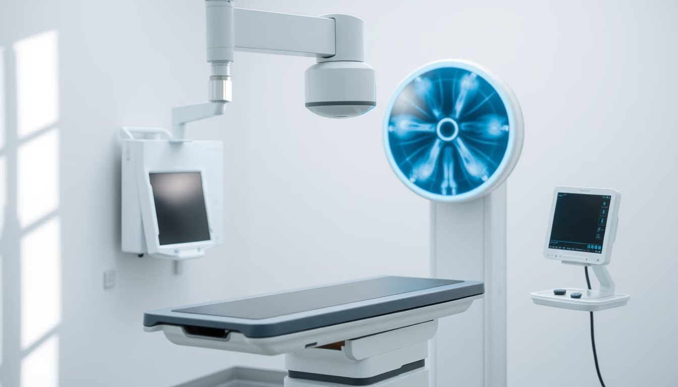
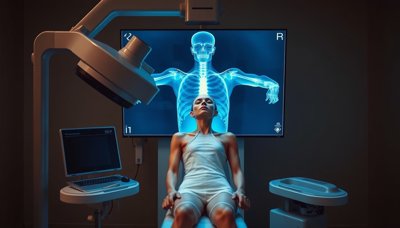
Orthopedic X-ray machine: essential equipment for diagnosing fractures and bone diseases, with digital technology.
About this subject
The orthopedic X-ray machine is a specialized medical device for imaging the skeletal system, including bones, joints, and surrounding tissues. Unlike conventional X-ray units, these machines are optimized to provide high resolution in specific areas such as hands, wrists, knees, and spine, assisting orthopedists in identifying fractures, dislocations, osteoarthritis, osteoporosis, and bone tumors. The exam is quick, non-invasive, and essential in emergency rooms and specialized clinics.
Technological evolution has transformed these machines: modern models use digital flat-panel detectors, replacing old radiographic films. This reduces radiation exposure time by up to 50%, improves image sharpness, and allows contrast adjustments without repeating the exam. Many units feature articulated arms and motorized positioning systems, facilitating examination of patients with reduced mobility. Furthermore, integration with PACS (Picture Archiving and Communication System) enables remote storage and sharing of images.
In Brazil, the National Health Surveillance Agency (ANVISA) regulates the use of these machines, requiring periodic calibration and operator training. Public and private hospitals increasingly invest in orthopedic X-ray rooms with adequate radiological protection, following standards from the National Nuclear Energy Commission (CNEN). Portability is also a trend: mobile units are used in ICUs, operating rooms, and even in field emergency care, such as sports events.
Interestingly, orthopedics was one of the first specialties to adopt radiography, shortly after Wilhelm Röntgen's discovery of X-rays in 1895. Since then, diagnostic imaging has become inseparable from orthopedic treatment. Studies show that for femoral fractures, plain radiography remains the gold standard for surgical decision-making, although CT and MRI complement complex cases. Patient safety is a priority: the effective dose of a typical orthopedic X-ray is equivalent to a few days of natural background radiation.
Frequently Asked Questions
What is an orthopedic X-ray machine used for?
It is used to diagnose fractures, dislocations, arthritis, bone tumors, and other musculoskeletal conditions by providing detailed images of bones and joints.
Is the orthopedic X-ray machine harmful to health?
It involves low radiation exposure, equivalent to a few days of natural background radiation. Modern machines further reduce doses, and strict safety protocols are followed.
How is an orthopedic X-ray different from a conventional X-ray?
Orthopedic X-rays are optimized for high-resolution imaging of specific skeletal areas, while conventional X-rays are used for general purposes like chest or abdomen.
Direct URL
https://pub-c7d6a6ea828543ac903a74a341ccb2e1.r2.dev/imagens/orthopedic-xray-machine-magazine-editorial-p2.avifHow to credit
Include a visible link back to UtilizAí. Copy one of the snippets below:
<a href="https://xn--utiliza-eza.com/en/midia/imagens/orthopedic-xray-machine-magazine-editorial-p2">Orthopedic xray machine</a> by <a href="https://xn--utiliza-eza.com">UtilizAí</a>, licensed under <a href="https://creativecommons.org/licenses/by/4.0/">CC BY 4.0</a>.
[Orthopedic xray machine](https://xn--utiliza-eza.com/en/midia/imagens/orthopedic-xray-machine-magazine-editorial-p2) by [UtilizAí](https://xn--utiliza-eza.com), CC BY 4.0
License: CC-BY-4.0