Orthopedic xray machine
1344×768 · AVIF · CC BY 4.0
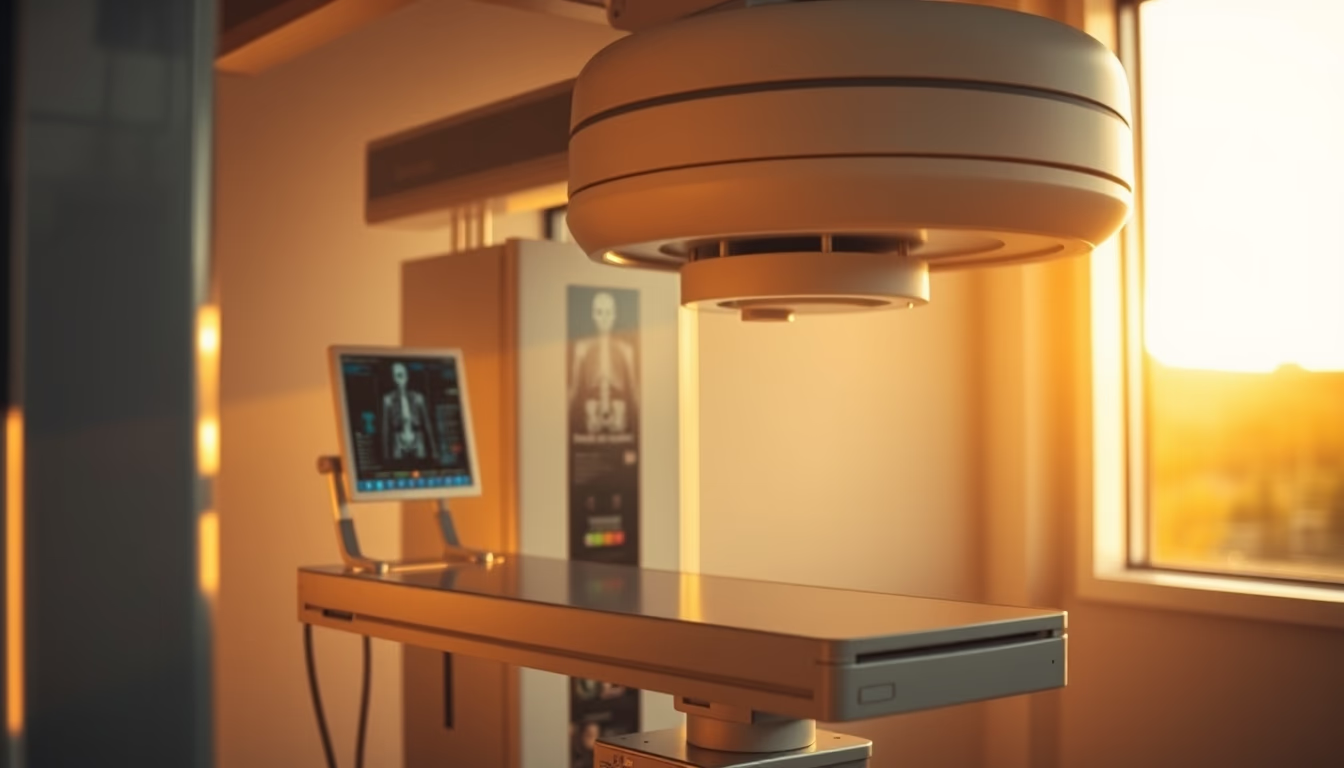
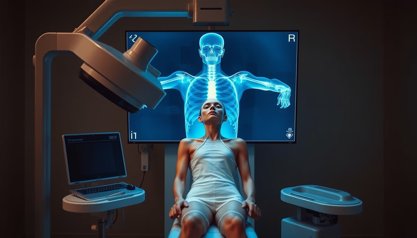
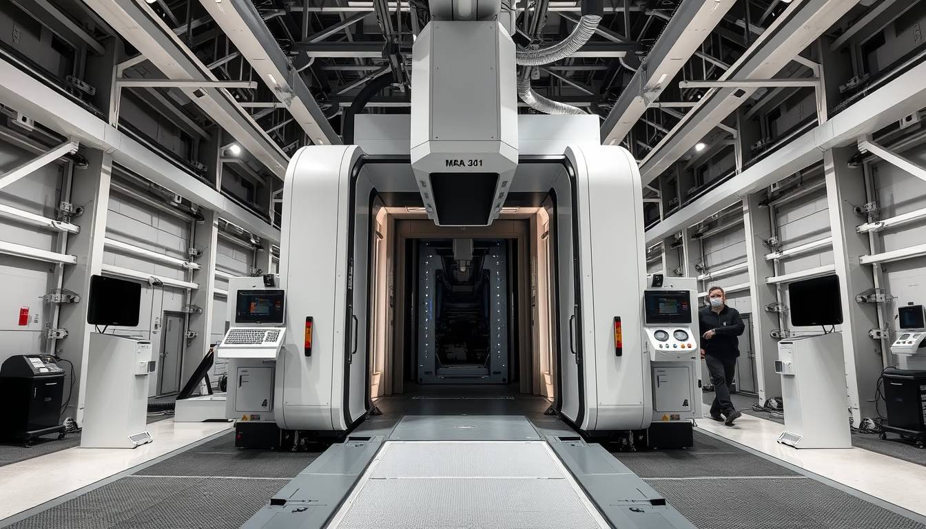
The orthopedic X-ray machine is a medical device essential for diagnosing fractures and bone diseases, used in clinics and hospitals.
About this subject
The orthopedic X-ray machine is designed to capture detailed images of the musculoskeletal system, especially bones and joints. It uses ionizing radiation to generate radiographs that reveal fractures, dislocations, osteoarthritis, osteoporosis, and bone tumors. Unlike CT scanners or MRIs, the orthopedic X-ray offers speed and low cost, making it the first-line investigation in orthopedic trauma.
Historically, clinical use of X-rays began shortly after Wilhelm Röntgen's discovery in 1895. In orthopedics, the technique became established in the following decades, allowing visualization of fractures without exploratory surgery. Modern equipment, such as C-arms and L-arms, facilitates patient positioning and beam angulation, reducing the need for repositioning. Digitalization replaced radiographic films with flat-panel detectors or CR (computed radiography), speeding up diagnosis and enabling contrast and zoom adjustments.
In many countries, including Brazil, the orthopedic X-ray machine is regulated by health agencies (e.g., ANVISA) and is present in both public (SUS) and private healthcare. Common exams include radiographs of the spine, hip, knee, wrist, and ankle. Radiation dose is low: a chest X-ray is about 0.1 mSv, while a lumbar spine X-ray can reach 1.5 mSv. Protective measures, such as lead aprons and beam collimation, are mandatory to minimize unnecessary exposure.
The equipment has also evolved with portable units used in ICUs and surgical centers, and with artificial intelligence software that aids in detecting subtle fractures. Preventive maintenance and periodic calibration are essential to ensure image quality and patient safety. Orthopedics, therefore, heavily relies on this accessible and effective technology.
Frequently Asked Questions
How long does an orthopedic X-ray exam take?
It usually takes 5 to 15 minutes, including patient positioning and image acquisition. Results are available within minutes after digital capture.
What are the radiation risks of an orthopedic X-ray?
The radiation dose is low and considered safe for adults. The risk is comparable to natural background exposure over a few days. Pregnant women should avoid the exam unless in emergencies with abdominal shielding.
Is any special preparation needed for an orthopedic X-ray?
No fasting or special preparation is required. It is recommended to wear comfortable clothing and remove metal objects such as jewelry and belts from the area to be examined.
Direct URL
https://pub-c7d6a6ea828543ac903a74a341ccb2e1.r2.dev/imagens/orthopedic-xray-machine-travel-photography-p4.avifHow to credit
Include a visible link back to UtilizAí. Copy one of the snippets below:
<a href="https://xn--utiliza-eza.com/en/midia/imagens/orthopedic-xray-machine-travel-photography-p4">Orthopedic xray machine</a> by <a href="https://xn--utiliza-eza.com">UtilizAí</a>, licensed under <a href="https://creativecommons.org/licenses/by/4.0/">CC BY 4.0</a>.
[Orthopedic xray machine](https://xn--utiliza-eza.com/en/midia/imagens/orthopedic-xray-machine-travel-photography-p4) by [UtilizAí](https://xn--utiliza-eza.com), CC BY 4.0
License: CC-BY-4.0