Orthopedic xray machine
1344×768 · AVIF · CC BY 4.0
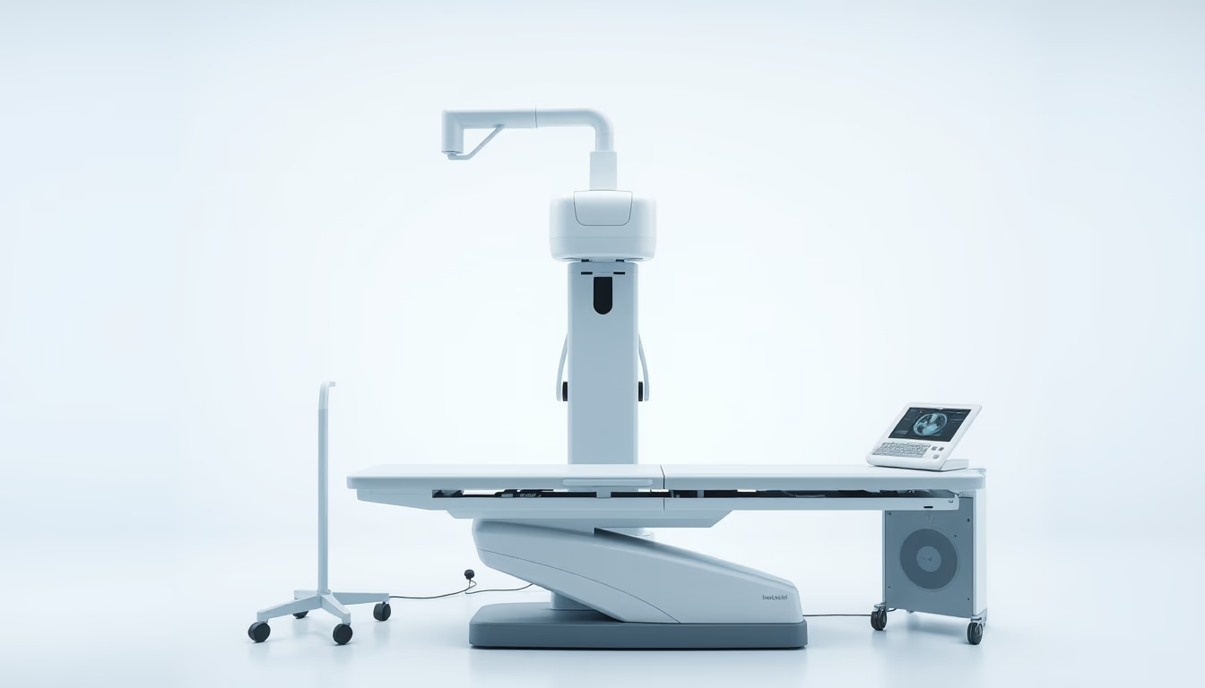
Orthopedic X-ray machine: essential equipment for diagnosing fractures and bone diseases, used in clinics and hospitals.
About this subject
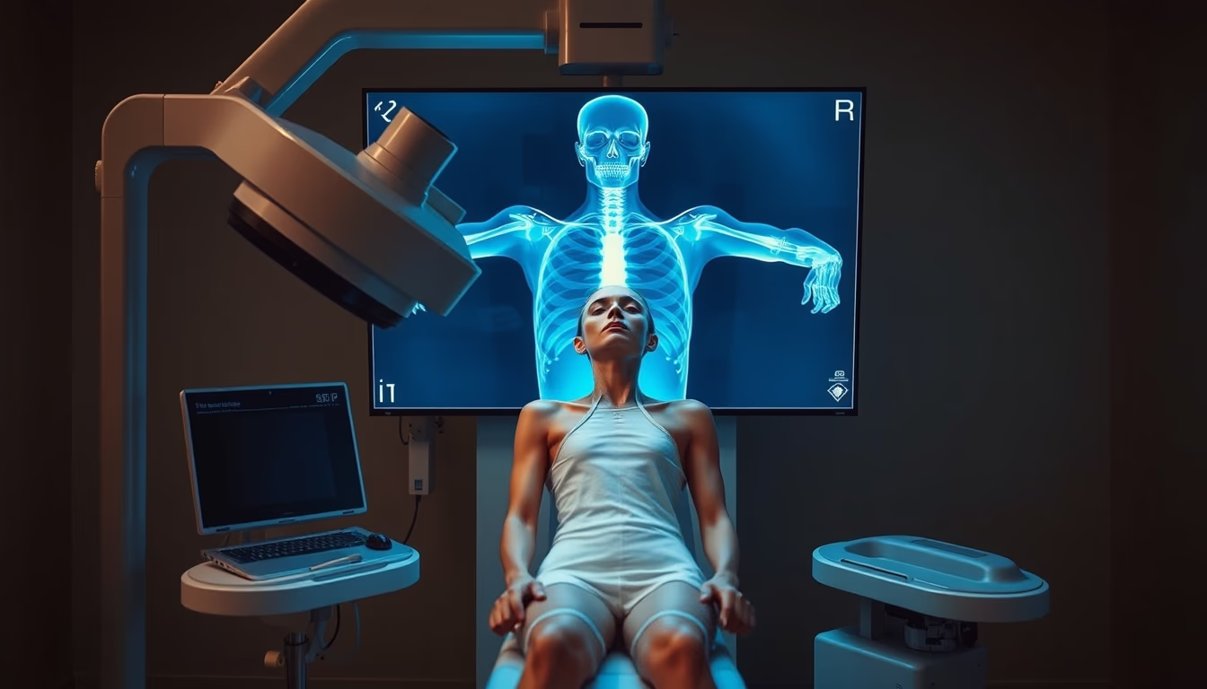
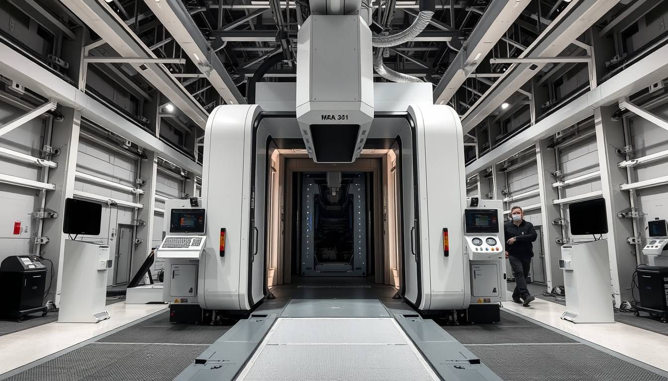
The orthopedic X-ray machine is a medical device specialized in imaging the musculoskeletal system, including bones, joints, and spine. Unlike conventional X-ray units, these devices are engineered to provide detailed views of fractures, dislocations, osteoarthritis, bone tumors, and deformities, assisting orthopedists in surgical planning and treatment monitoring. Technology ranges from analog to digital systems, with high-resolution sensors that lower radiation exposure without compromising image quality.
In the United States, the use of orthopedic X-rays follows guidelines from the American College of Radiology, specifying standard projections such as anterior-posterior and lateral views for upper and lower extremities. The U.S. Food and Drug Administration (FDA) regulates equipment safety and calibration, ensuring radiation protection. Approximately 80% of orthopedic diagnoses initially rely on plain radiographs, making this machine indispensable in outpatient and emergency settings.
Interestingly, modern orthopedic X-ray machines deliver doses as low as 0.1 mSv per examination, equivalent to about 10 days of natural background radiation. Advances such as intraoperative fluoroscopy allow surgeons to see implant placement in real time during procedures like fracture fixation. The development of portable and C-arm systems has expanded their use into clinics and intensive care units, where immobile patients can be examined without transport.
Frequently Asked Questions
What is the difference between an orthopedic X-ray and a conventional one?
Orthopedic X-rays are optimized for bone and joint visualization, with specific kVp and mAs settings, plus tighter collimation to reduce scatter. Modern equipment includes image processing algorithms that enhance bone edges.
How many orthopedic X-ray exams are safe per year?
There is no absolute limit, as each exam has a low dose (about 0.1 mSv). For reference, annual background exposure is about 3 mSv. The physician evaluates risk-benefit, especially in children and pregnant women.
How does a digital orthopedic X-ray system work?
Instead of film, a digital detector (DR or CR) captures the image. Sensors convert X-ray photons into an electrical signal, processed and displayed on a computer in seconds. This allows contrast and brightness adjustments without repeating exams.
Direct URL
https://pub-c7d6a6ea828543ac903a74a341ccb2e1.r2.dev/imagens/orthopedic-xray-machine-commercial-product-photography-p3.avifHow to credit
Include a visible link back to UtilizAí. Copy one of the snippets below:
<a href="https://xn--utiliza-eza.com/en/midia/imagens/orthopedic-xray-machine-commercial-product-photography-p3">Orthopedic xray machine</a> by <a href="https://xn--utiliza-eza.com">UtilizAí</a>, licensed under <a href="https://creativecommons.org/licenses/by/4.0/">CC BY 4.0</a>.
[Orthopedic xray machine](https://xn--utiliza-eza.com/en/midia/imagens/orthopedic-xray-machine-commercial-product-photography-p3) by [UtilizAí](https://xn--utiliza-eza.com), CC BY 4.0
License: CC-BY-4.0