Mri machine gantry
1344×768 · AVIF · CC BY 4.0

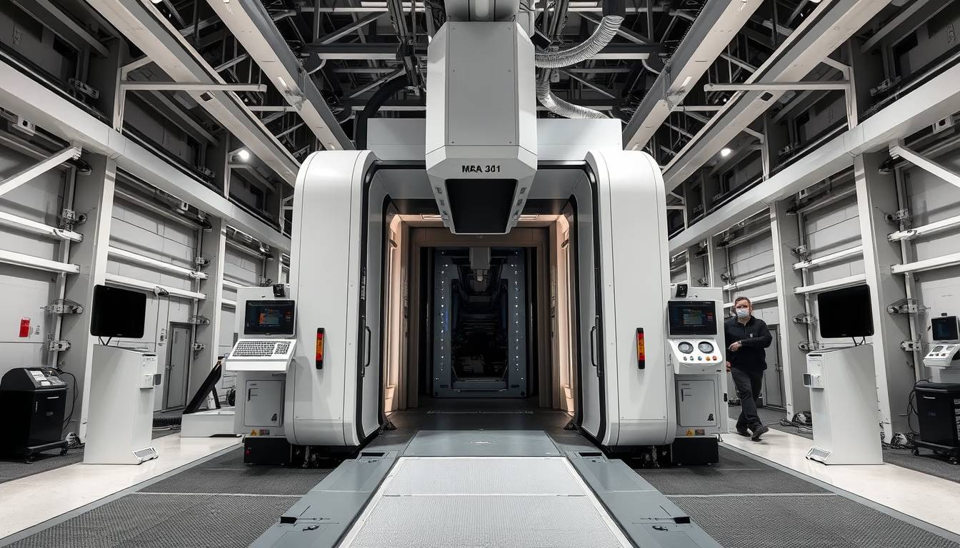
The gantry of an MRI machine is the ring-shaped structure that houses key components for generating high-resolution images of the human body.
About this subject
The gantry is the most iconic component of a magnetic resonance imaging (MRI) machine. It is a large superconducting magnet shaped like a ring, typically with an inner diameter of 60 to 70 centimeters, that generates a strong, uniform magnetic field. Inside the gantry are also the gradient coils and radiofrequency coils, which spatially encode the signal and excite the protons in the tissue. When the patient is positioned at the center of the gantry, hydrogen protons in the body align with the magnetic field and, after radiofrequency pulses, emit signals that are captured and transformed into detailed images.
MRI was developed based on the work of Paul Lauterbur and Peter Mansfield, who received the Nobel Prize in Physiology or Medicine in 2003. The first commercial scanner appeared in the early 1980s. Since then, the technology has evolved significantly: magnetic field strengths increased from 0.5 Tesla (T) to 1.5T and 3T in clinical practice, and up to 7T in research. The gantry itself has become shorter and wider (wide-bore), reducing claustrophobia and allowing exams in obese patients or those with specific implants. Open MRI systems with partial gantries are also available for greater comfort.
The main advantage of MRI is its excellent soft-tissue contrast, essential for neurological, musculoskeletal, and oncological diagnoses. Unlike computed tomography, it uses no ionizing radiation. Thus, the gantry is not merely a housing; it concentrates the precision engineering that enables one of the most important imaging methods in modern medicine.
Frequently Asked Questions
What is the difference between the gantry and the MRI scanner?
The gantry is the part of the scanner that contains the main magnet, gradient coils, and radiofrequency coils. The scanner also includes the patient table, control console, and reconstruction computer. In short, the gantry is the ring where the patient is inserted.
How does MRI work inside the gantry?
Inside the gantry, a strong magnetic field aligns the body's hydrogen protons. Radiofrequency pulses then excite them. As they relax, they emit signals that are captured by coils and used to form images based on tissue density and properties.
What do the magnetic field strengths of 1.5T and 3T mean in MRI?
Tesla (T) is the unit of magnetic field strength. 1.5T is the clinical standard for most exams, offering a good balance between image quality and safety. 3T provides higher signal-to-noise ratio, allowing more detailed images and faster scans, but requires more care with artifacts.
Direct URL
https://pub-c7d6a6ea828543ac903a74a341ccb2e1.r2.dev/imagens/mri-machine-gantry-high-fashion-editorial-p5.avifHow to credit
Include a visible link back to UtilizAí. Copy one of the snippets below:
<a href="https://xn--utiliza-eza.com/en/midia/imagens/mri-machine-gantry-high-fashion-editorial-p5">Mri machine gantry</a> by <a href="https://xn--utiliza-eza.com">UtilizAí</a>, licensed under <a href="https://creativecommons.org/licenses/by/4.0/">CC BY 4.0</a>.
[Mri machine gantry](https://xn--utiliza-eza.com/en/midia/imagens/mri-machine-gantry-high-fashion-editorial-p5) by [UtilizAí](https://xn--utiliza-eza.com), CC BY 4.0
License: CC-BY-4.0