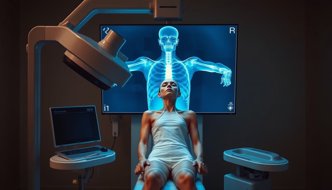
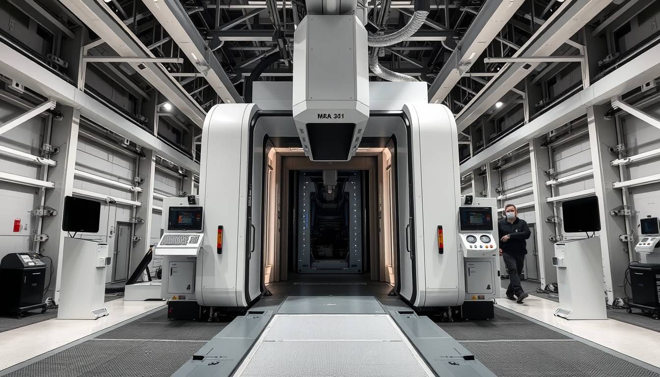
Orthopedic xray machine
1344×768 · AVIF · CC BY 4.0
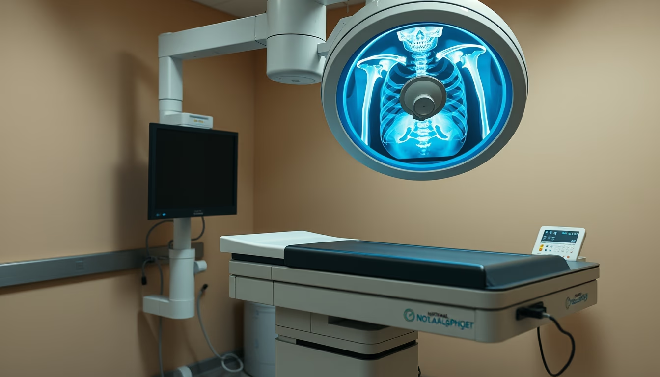
Orthopedic X-ray machine vital for diagnosing fractures and bone diseases, providing detailed images of limbs and joints.
About this subject
The orthopedic X-ray machine is an essential tool in medicine, particularly in orthopedics and traumatology. Unlike conventional X-ray devices, these units are optimized to visualize bones, joints, and adjacent soft tissues with high resolution. They enable diagnosis of fractures, dislocations, osteoporosis, arthritis, and other conditions affecting the musculoskeletal system. In emergency settings, rapid image acquisition is critical for timely treatment decisions.
Operation relies on ionizing radiation that passes through the patient's body. Bone tissue absorbs more radiation, appearing white on images, while soft tissues appear in gray tones. Modern detectors, such as digital flat panels, convert X-rays into electrical signals to produce high-quality images. Many orthopedic machines feature articulated arms (C-arm) that allow variable angles, essential for lateral or oblique views. During surgical procedures, real-time fluoroscopy guides the placement of pins and screws.
Technological advances have brought significant benefits: reduced radiation dose, instant image processing, and integration with Picture Archiving and Communication Systems (PACS). Portable devices have become common in orthopedics clinics, increasing diagnostic accessibility. In Brazil, the National Health Surveillance Agency (ANVISA) regulates these devices to ensure safety and quality. Recent studies highlight the role of artificial intelligence in automatically interpreting orthopedic X-rays, speeding up diagnoses and reducing human errors.
Frequently Asked Questions
What distinguishes an orthopedic X-ray from a standard X-ray?
Orthopedic X-rays are tailored for limbs and joints, with specific exposure settings to enhance bone structures. Many units feature C-arms for variable angles, especially useful during surgeries.
Is it safe to have multiple orthopedic X-ray exams?
Yes, as long as the radiation dose is controlled and clinically justified. Modern machines use low doses, but unnecessary exposure should be avoided, especially in children and pregnant women.
How does the image acquisition process work?
The patient is positioned between the X-ray source and detector. Rays pass through the body, and differential attenuation by tissues creates the image. Digital detectors convert rays into electrical signals for immediate viewing.
Direct URL
https://pub-c7d6a6ea828543ac903a74a341ccb2e1.r2.dev/imagens/orthopedic-xray-machine-national-geographic-style-p5.avifHow to credit
Include a visible link back to UtilizAí. Copy one of the snippets below:
<a href="https://xn--utiliza-eza.com/en/midia/imagens/orthopedic-xray-machine-national-geographic-style-p5">Orthopedic xray machine</a> by <a href="https://xn--utiliza-eza.com">UtilizAí</a>, licensed under <a href="https://creativecommons.org/licenses/by/4.0/">CC BY 4.0</a>.
[Orthopedic xray machine](https://xn--utiliza-eza.com/en/midia/imagens/orthopedic-xray-machine-national-geographic-style-p5) by [UtilizAí](https://xn--utiliza-eza.com), CC BY 4.0
License: CC-BY-4.0