Orthopedic xray machine
1344×768 · AVIF · CC BY 4.0
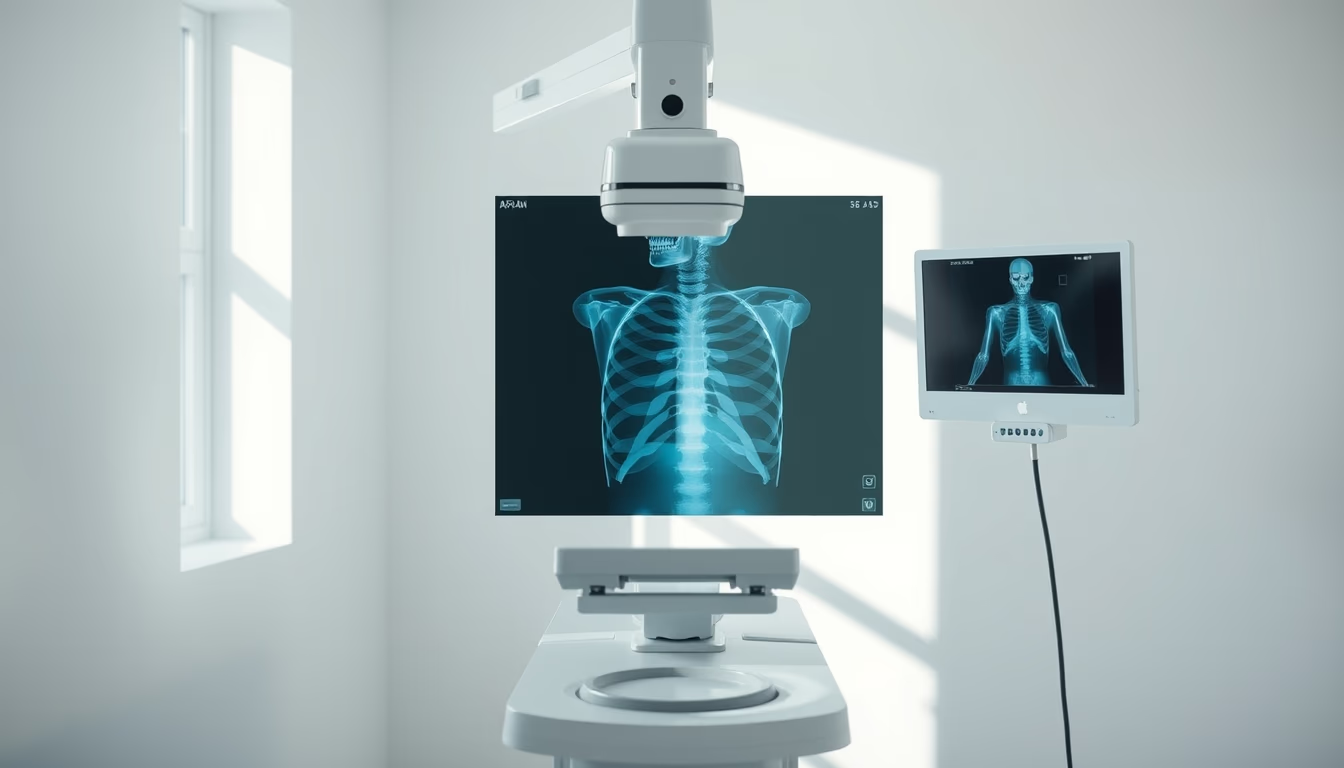
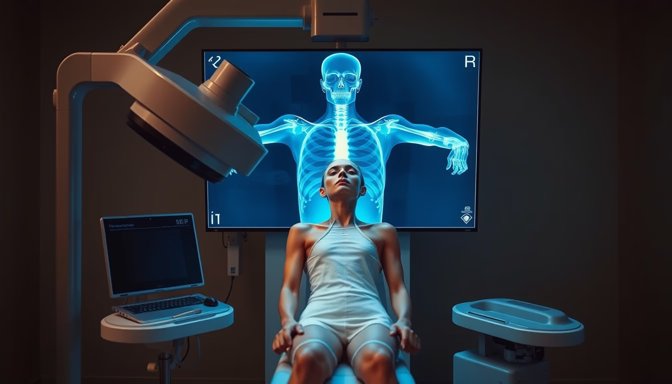
An orthopedic X-ray machine is essential for diagnosing fractures and bone diseases, using ionizing radiation to produce detailed images.
About this subject
An orthopedic X-ray machine is a medical device specifically designed to capture images of the musculoskeletal system, including bones, joints, and adjacent tissues. Unlike general radiology units, many orthopedic models are compact and adjustable, allowing precise positioning for extremities such as hands, feet, knees, and the spine. The operating principle is based on the emission of X-rays that pass through the body and are attenuated differently by dense structures (bones) and soft tissues, producing a grayscale image.
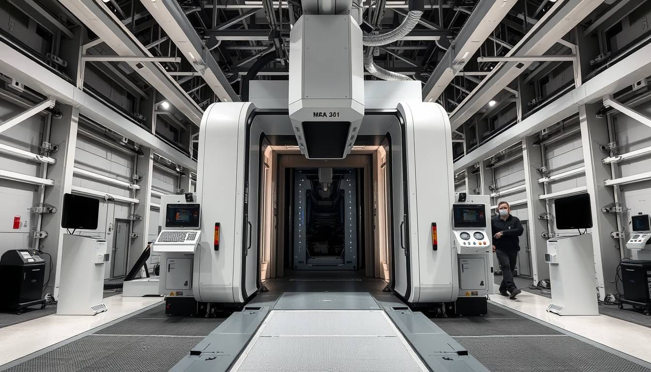
In clinical practice, orthopedic X-ray is the first-line tool for evaluating fractures, dislocations, osteoarthritis, osteoporosis, and bone tumors. Standard views such as anteroposterior and lateral are used to assess bone alignment and healing. With technological advances, digital radiography (DR) systems have replaced film-based radiography, offering higher resolution, lower radiation exposure, and instant image processing. Equipment like the C-arm or mini C-arm is used in orthopedic surgeries to guide procedures such as fracture fixation and joint arthroplasty.
Radiation safety is a priority: operators wear lead aprons and dosimeters, and the dose is kept as low as reasonably achievable (ALARA principle). In Brazil, the National Health Surveillance Agency (ANVISA) regulates the use of X-ray equipment, requiring licensing and quality control. Modern orthopedic machines may include features such as automatic exposure control, collimation, and image processing to enhance diagnostic accuracy.
Frequently Asked Questions
How does an orthopedic X-ray machine work?
It emits X-rays that pass through the body. Bones, being dense, absorb more radiation and appear white on the image, while soft tissues appear in shades of gray. A detector (digital or film) captures the remaining radiation to form the image.
What are the risks of radiation in orthopedic exams?
Exposure to ionizing radiation carries a theoretical cancer risk, but doses in orthopedic exams are low (e.g., a chest X-ray equals about 10 days of natural background radiation). Protective measures like lead aprons and collimation minimize risks.
What is the difference between digital and conventional X-ray?
Digital X-ray uses electronic sensors to convert X-rays into digital signals, allowing immediate viewing on a computer, contrast adjustment, and storage. Conventional X-ray uses chemical film that requires processing and cannot be post-processed.
Direct URL
https://pub-c7d6a6ea828543ac903a74a341ccb2e1.r2.dev/imagens/orthopedic-xray-machine-minimalist-composition-p0.avifHow to credit
Include a visible link back to UtilizAí. Copy one of the snippets below:
<a href="https://xn--utiliza-eza.com/en/midia/imagens/orthopedic-xray-machine-minimalist-composition-p0">Orthopedic xray machine</a> by <a href="https://xn--utiliza-eza.com">UtilizAí</a>, licensed under <a href="https://creativecommons.org/licenses/by/4.0/">CC BY 4.0</a>.
[Orthopedic xray machine](https://xn--utiliza-eza.com/en/midia/imagens/orthopedic-xray-machine-minimalist-composition-p0) by [UtilizAí](https://xn--utiliza-eza.com), CC BY 4.0
License: CC-BY-4.0