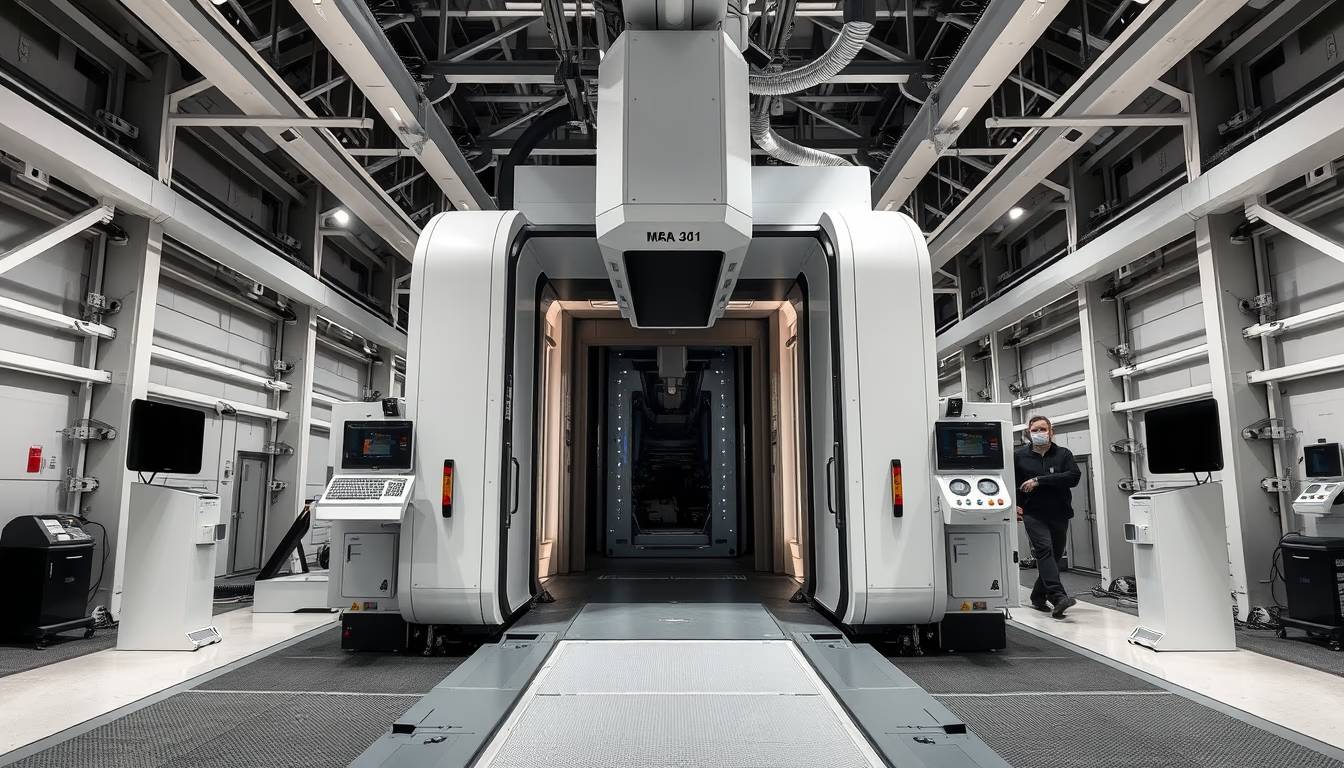
Mri machine gantry
1344×768 · AVIF · CC BY 4.0

The gantry of an MRI machine is the ring-shaped structure that houses the superconducting magnet and gradient coils, essential for acquiring high-resolution medical images.
About this subject
The gantry of an MRI machine is the most iconic part of the device: a cylindrical tunnel typically 60 to 70 centimeters in diameter that houses the superconducting magnet. This magnet, cooled to near absolute zero using liquid helium, generates a strong static magnetic field, usually between 1.5 and 3 Tesla in standard clinical machines. Inside the gantry are gradient coils, which spatially encode proton signals from body tissues, and radiofrequency coils that excite and receive signals to form images.
The gantry's architecture directly affects patient experience: closed-bore models can induce claustrophobia, while open or wide-bore systems with larger or vertical designs have been developed to reduce discomfort. Advances include parallel imaging sequences (e.g., GRAPPA or SENSE) using multiple receiver coils placed in the gantry for faster scans and less noise. Research centers use 7 Tesla or higher machines with larger gantries and specialized acoustic insulation for neurological and preclinical studies.
Historically, the first full clinical MRI scanner was installed in 1984 at the University of Aberdeen, UK, with a much simpler and bulkier gantry than today's models. Current manufacturers like Siemens, GE Healthcare, and Philips compete to shorten gantry length and enlarge bore diameter, alleviating confinement. Some recent models offer 70 cm or larger bores and tunnel lengths of just 125 cm, with embedded lighting and silent ventilation for comfort.
Beyond clinical use, gantry design affects applications such as intraoperative MRI, where the machine is installed in surgical suites, and whole-body MRI, requiring uniform magnetic fields over a large volume. Maintenance involves periodic liquid helium refills (cryogen refill) and coil calibration, critical for image quality. From an artistic perspective, fine art photography of the gantry explores its geometric shapes, symmetry, and the contrast between cold technology and the hospital setting, highlighting the equipment's inherent beauty.
Frequently Asked Questions
What is the gantry of an MRI machine?
It is the ring-shaped structure that contains the main magnet, gradient coils, and radiofrequency coils. It is the part where the patient is positioned for the scan.
Why do some patients feel claustrophobia inside the gantry?
Because the tunnel is narrow (60-70 cm in diameter) and enclosed, which can trigger anxiety. Open-bore or wider-bore machines (70+ cm) help reduce this discomfort.
What is the difference between a 1.5T and a 3T gantry?
The main difference is magnetic field strength: 3T provides higher signal-to-noise ratio and resolution, enabling faster or more detailed scans. However, it may increase artifacts and tissue heating, requiring optimized sequences.
Direct URL
https://pub-c7d6a6ea828543ac903a74a341ccb2e1.r2.dev/imagens/mri-machine-gantry-fine-art-photography-p0.avifHow to credit
Include a visible link back to UtilizAí. Copy one of the snippets below:
<a href="https://xn--utiliza-eza.com/en/midia/imagens/mri-machine-gantry-fine-art-photography-p0">Mri machine gantry</a> by <a href="https://xn--utiliza-eza.com">UtilizAí</a>, licensed under <a href="https://creativecommons.org/licenses/by/4.0/">CC BY 4.0</a>.
[Mri machine gantry](https://xn--utiliza-eza.com/en/midia/imagens/mri-machine-gantry-fine-art-photography-p0) by [UtilizAí](https://xn--utiliza-eza.com), CC BY 4.0
License: CC-BY-4.0