Protein structure 3d
1344×768 · AVIF · CC BY 4.0
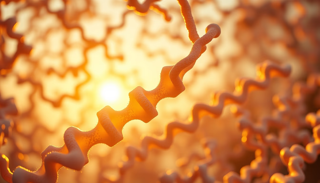
A estrutura tridimensional de proteínas determina sua função biológica e é alvo de estudos por cristalografia de raios X e criomicroscopia eletrônica.
Sobre o tema
Proteínas são macromoléculas essenciais para a vida, desempenhando funções como catálise enzimática, transporte de moléculas e sinalização celular. A sua atividade depende diretamente do dobramento tridimensional, ou conformação nativa, que é estabilizada por interações não covalentes, incluindo pontes de hidrogênio, interações hidrofóbicas e ligações iônicas. Métodos experimentais como a cristalografia de raios X, a ressonância magnética nuclear (RMN) e a criomicroscopia eletrônica (crio-EM) permitem determinar estruturas atômicas de proteínas, contribuindo para o entendimento de doenças e o desenvolvimento de fármacos.
A primeira estrutura de proteína resolvida em nível atômico foi a da mioglobina, por John Kendrew em 1958, utilizando cristalografia de raios X. Desde então, o Protein Data Bank (PDB) acumula mais de 200 mil estruturas depositadas, disponíveis gratuitamente para a comunidade científica. A crio-EM, reconhecida com o Prêmio Nobel de Química em 2017, revolucionou a área ao permitir a visualização de proteínas grandes e complexos macromoleculares sem necessidade de cristalização.
O dobramento incorreto de proteínas está associado a patologias como Alzheimer, Parkinson e fibrose cística. Compreender a estrutura tridimensional ajuda a projetar inibidores ou moduladores alostéricos que interagem com sítios específicos, uma abordagem central na química medicinal moderna. Além disso, técnicas computacionais como docking molecular e dinâmica molecular simulam interações ligante-proteína, acelerando a descoberta de novos candidatos a medicamentos.
Perguntas frequentes
O que é o Protein Data Bank (PDB)?
O PDB é um banco de dados internacional que armazena estruturas tridimensionais de proteínas e ácidos nucleicos determinadas experimentalmente. É acessível gratuitamente e usado por pesquisadores para estudos estruturais e desenvolvimento de fármacos.
Como a estrutura 3D de uma proteína é determinada?
As principais técnicas são cristalografia de raios X, ressonância magnética nuclear (RMN) e criomicroscopia eletrônica (crio-EM). Cada uma tem vantagens e limitações, e a escolha depende do tamanho e da natureza da proteína.
Por que a estrutura tridimensional é importante para a medicina?
Conhecer a estrutura permite identificar sítios ativos e alostéricos, essenciais para o desenho racional de medicamentos. Também ajuda a entender mutações que causam doenças e a desenvolver terapias direcionadas.
URL direta
https://pub-c7d6a6ea828543ac903a74a341ccb2e1.r2.dev/imagens/protein-structure-3d-cinematic-wide-shot-p0.avifComo creditar
Inclua um link visível de volta pro UtilizAí. Copie um dos snippets abaixo:
<a href="https://xn--utiliza-eza.com/midia/imagens/protein-structure-3d-cinematic-wide-shot-p0">Protein structure 3d</a> by <a href="https://xn--utiliza-eza.com">UtilizAí</a>, licensed under <a href="https://creativecommons.org/licenses/by/4.0/">CC BY 4.0</a>.
[Protein structure 3d](https://xn--utiliza-eza.com/midia/imagens/protein-structure-3d-cinematic-wide-shot-p0) by [UtilizAí](https://xn--utiliza-eza.com), CC BY 4.0
Licença: CC-BY-4.0





