Nucleus chromosomes microscope
1344×768 · AVIF · CC BY 4.0
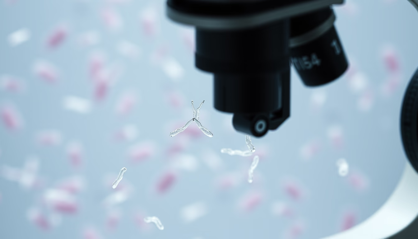
Imagem de núcleo celular com cromossomos visíveis ao microscópio, evidenciando a estrutura do DNA durante a divisão celular.
Sobre o tema
O núcleo celular é uma organela fundamental encontrada em células eucarióticas, abrigando a maior parte do material genético na forma de cromossomos. Durante a interfase, os cromossomos estão descondensados como cromatina, mas durante a mitose ou meiose tornam-se visíveis como estruturas espessas e enroladas. A microscopia óptica, especialmente com corantes como DAPI ou Giemsa, permite visualizar essas estruturas, sendo essencial para estudos de citogenética e diagnóstico de anomalias cromossômicas.
Os cromossomos humanos consistem em 23 pares, incluindo um par de cromossomos sexuais. Técnicas de bandeamento, como bandas G, revelam padrões característicos que ajudam a identificar rearranjos como translocações e deleções, associados a doenças genéticas. A microscopia de fluorescência, combinada com sondas de DNA (FISH), permite localizar sequências específicas, refinando o diagnóstico.
Além do valor clínico, o estudo dos cromossomos fornece insights sobre evolução e biologia molecular. A descoberta da estrutura helicoidal do DNA por Watson e Crick em 1953 baseou-se em dados de difração de raios X, mas a visualização direta de cromossomos ao microscópio continua sendo uma ferramenta didática e de pesquisa. Imagens como esta representam a interseção entre observação clássica e técnicas modernas de biologia celular.
Perguntas frequentes
O que são cromossomos e por que são visíveis apenas durante a divisão celular?
Cromossomos são estruturas formadas por DNA e proteínas (histonas) que carregam os genes. Durante a maior parte do ciclo celular (interfase), eles estão descondensados para permitir a transcrição gênica, mas durante a mitose ou meiose eles se condensam para facilitar a distribuição igualitária do material genético entre as células-filhas, tornando-se visíveis ao microscópio óptico.
Quais técnicas de coloração são usadas para visualizar cromossomos?
As técnicas mais comuns incluem o corante DAPI (que se liga especificamente ao DNA) e a coloração Giemsa (usada em bandeamento G, que produz padrões de bandas claras e escuras). A hibridização in situ fluorescente (FISH) usa sondas marcadas para detectar sequências específicas de DNA. Para análise de cariótipo, geralmente utiliza-se bandeamento G ou C.
Qual a importância clínica da análise de cromossomos?
A análise cromossômica é fundamental para diagnosticar síndromes genéticas como a síndrome de Down (trissomia do 21), de Turner (monossomia do X) e de Klinefelter (XXY). Também identifica translocações associadas a leucemias e outros cânceres, orientando o prognóstico e tratamento. A citogenética é uma ferramenta padrão em medicina fetal e reprodutiva.
URL direta
https://pub-c7d6a6ea828543ac903a74a341ccb2e1.r2.dev/imagens/nucleus-chromosomes-microscope-magazine-editorial-p4.avifComo creditar
Inclua um link visível de volta pro UtilizAí. Copie um dos snippets abaixo:
<a href="https://xn--utiliza-eza.com/midia/imagens/nucleus-chromosomes-microscope-magazine-editorial-p4">Nucleus chromosomes microscope</a> by <a href="https://xn--utiliza-eza.com">UtilizAí</a>, licensed under <a href="https://creativecommons.org/licenses/by/4.0/">CC BY 4.0</a>.
[Nucleus chromosomes microscope](https://xn--utiliza-eza.com/midia/imagens/nucleus-chromosomes-microscope-magazine-editorial-p4) by [UtilizAí](https://xn--utiliza-eza.com), CC BY 4.0
Licença: CC-BY-4.0





