Electron microscope sample
1344×768 · AVIF · CC BY 4.0
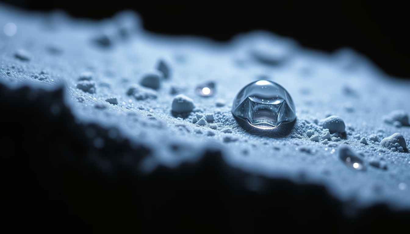
Amostras para microscopia eletrônica são preparadas com técnicas precisas para revelar estruturas em escala nanométrica, essenciais em biologia e ciência dos materiais.
Sobre o tema
Amostras para microscopia eletrônica passam por um processo rigoroso de preparação para suportar o vácuo e o feixe de elétrons. Em biologia, tecidos são fixados com glutaraldeído, desidratados em álcool e embebidos em resina. Cortes ultrafinos (50-100 nm) são obtidos com ultramicrótomo e depositados em grades de cobre. Para evitar acúmulo de carga, recobre-se a amostra com uma fina camada de ouro ou carbono, processo chamado metalização. Já em materiais, amostras sólidas precisam ser polidas ou atacadas quimicamente para revelar grãos e defeitos.
A microscopia eletrônica revolucionou a ciência ao permitir a visualização de vírus, organelas celulares e nanomateriais. O microscópio eletrônico de transmissão (TEM) utiliza elétrons que atravessam a amostra, gerando imagens com resolução subnanométrica. Já o microscópio de varredura (SEM) escaneia a superfície, produzindo imagens tridimensionais de alta profundidade de foco. Técnicas modernas incluem a criomicroscopia eletrônica, que congela amostras rapidamente para preservar estruturas nativas, Prêmio Nobel de Química em 2017.
Curiosidade: a primeira imagem de microscopia eletrônica foi obtida em 1931 por Ernst Ruska e Max Knoll, mostrando uma grade de metal com ampliação de 400 vezes. Hoje, a resolução chega a 0,05 nm, suficiente para visualizar átomos individuais. No Brasil, instituições como o LNLS e o INMETRO mantêm laboratórios de ponta para pesquisa e inovação.
Perguntas frequentes
Como as amostras biológicas são preparadas para microscopia eletrônica?
As amostras são fixadas quimicamente, desidratadas, incluídas em resina e cortadas em fatias ultrafinas com ultramicrótomo. Depois, são montadas em grades e metalizadas com ouro ou carbono para conduzir elétrons.
Qual a diferença entre microscópio eletrônico de varredura e transmissão?
O microscópio de transmissão (TEM) analisa elétrons que atravessam a amostra, revelando estrutura interna com altíssima resolução. O de varredura (SEM) escaneia a superfície, gerando imagens tridimensionais da topografia.
Por que as amostras precisam ser metalizadas?
A metalização com ouro ou carbono evita o acúmulo de carga elétrica na superfície da amostra, que distorceria o feixe de elétrons e degradaria a imagem. Também aumenta a emissão de elétrons secundários no SEM.
URL direta
https://pub-c7d6a6ea828543ac903a74a341ccb2e1.r2.dev/imagens/electron-microscope-sample-magazine-editorial-p8.avifComo creditar
Inclua um link visível de volta pro UtilizAí. Copie um dos snippets abaixo:
<a href="https://xn--utiliza-eza.com/midia/imagens/electron-microscope-sample-magazine-editorial-p8">Electron microscope sample</a> by <a href="https://xn--utiliza-eza.com">UtilizAí</a>, licensed under <a href="https://creativecommons.org/licenses/by/4.0/">CC BY 4.0</a>.
[Electron microscope sample](https://xn--utiliza-eza.com/midia/imagens/electron-microscope-sample-magazine-editorial-p8) by [UtilizAí](https://xn--utiliza-eza.com), CC BY 4.0
Licença: CC-BY-4.0





