Electron microscope sample
1344×768 · AVIF · CC BY 4.0
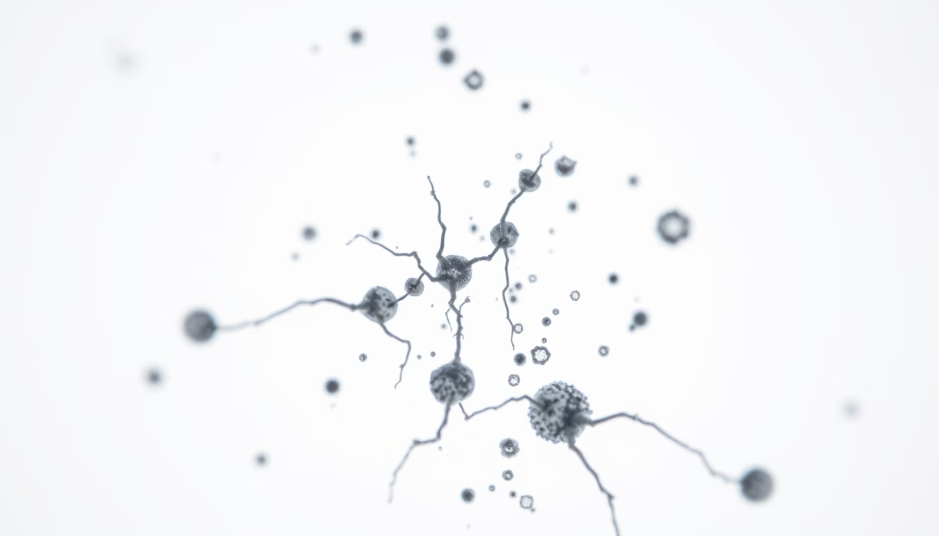
Amostras para microscopia eletrônica são preparadas com técnicas rigorosas para revelar estruturas celulares e moleculares em nanoescala.
Sobre o tema
A microscopia eletrônica é uma ferramenta fundamental na biologia moderna, permitindo a visualização de estruturas que vão desde organelas celulares até vírus e macromoléculas. Para que uma amostra seja observada em um microscópio eletrônico de varredura (SEM) ou de transmissão (TEM), ela precisa passar por um processo meticuloso de preparação. Isso inclui fixação química para preservar a estrutura, desidratação, embebição em resinas, corte ultrafino com ultramicrótomo e, no caso do SEM, metalização com ouro ou carbono para tornar a superfície condutora. Cada etapa é crítica para evitar artefatos e garantir que a imagem obtida represente fielmente a morfologia original. No Brasil, laboratórios de microscopia eletrônica estão presentes em universidades como a USP, Unicamp e UFRJ, além de centros de pesquisa como o LNLS (Laboratório Nacional de Luz Síncrotron). A técnica revolucionou áreas como a virologia, onde foi possível observar pela primeira vez partículas virais como o HIV, e a neurobiologia, com a reconstrução tridimensional de sinapses. Curiosamente, a preparação de amostras para microscopia eletrônica pode levar dias ou até semanas, dependendo da complexidade e do tipo de análise desejada.
Perguntas frequentes
Qual a diferença entre microscopia eletrônica de varredura (SEM) e de transmissão (TEM)?
No SEM, um feixe de elétrons varre a superfície da amostra, produzindo imagens tridimensionais da topografia. No TEM, os elétrons atravessam a amostra, gerando imagens bidimensionais de estruturas internas com resolução atômica.
Por que as amostras precisam ser metalizadas para SEM?
A metalização com ouro ou carbono torna a superfície da amostra condutora, evitando o acúmulo de cargas elétricas que distorceriam a imagem. Além disso, aumenta a emissão de elétrons secundários, melhorando o contraste.
Quanto tempo leva para preparar uma amostra para microscopia eletrônica?
O processo pode levar de algumas horas a várias semanas, dependendo da complexidade da amostra. Tecidos biológicos requerem fixação, desidratação, inclusão em resina e cortes ultrafinos, cada etapa demorada.
URL direta
https://pub-c7d6a6ea828543ac903a74a341ccb2e1.r2.dev/imagens/electron-microscope-sample-commercial-product-photography-p9.avifComo creditar
Inclua um link visível de volta pro UtilizAí. Copie um dos snippets abaixo:
<a href="https://xn--utiliza-eza.com/midia/imagens/electron-microscope-sample-commercial-product-photography-p9">Electron microscope sample</a> by <a href="https://xn--utiliza-eza.com">UtilizAí</a>, licensed under <a href="https://creativecommons.org/licenses/by/4.0/">CC BY 4.0</a>.
[Electron microscope sample](https://xn--utiliza-eza.com/midia/imagens/electron-microscope-sample-commercial-product-photography-p9) by [UtilizAí](https://xn--utiliza-eza.com), CC BY 4.0
Licença: CC-BY-4.0





