Cell division microscope
1344×768 · AVIF · CC BY 4.0
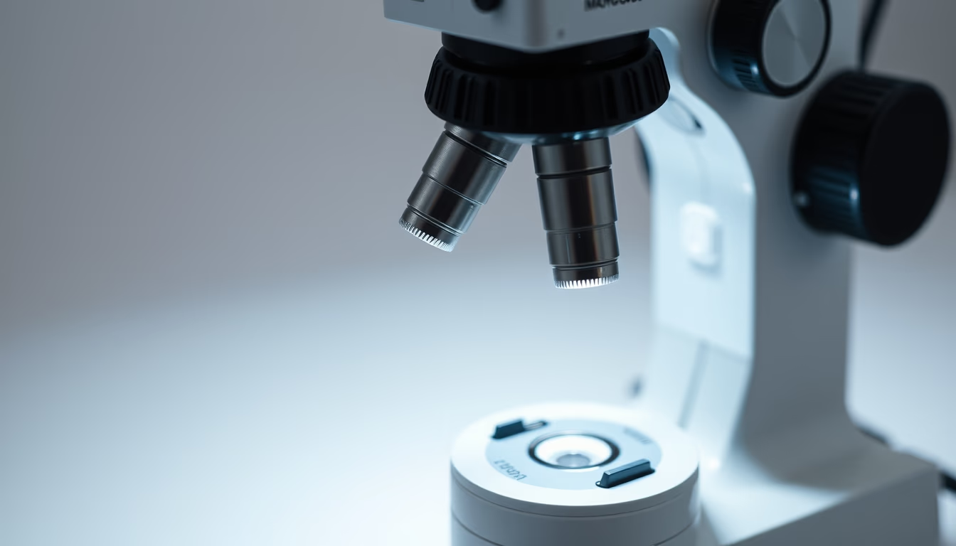
Cell division is the process by which cells reproduce, essential for growth and tissue repair. Under a microscope, the phases of mitosis and meiosis can be observed.
About this subject
Cell division is a fundamental process for life. In multicellular organisms, such as humans, it allows growth, tissue renewal, and reproduction. There are two main types: mitosis and meiosis. Mitosis produces two genetically identical daughter cells, crucial for development and repair. Meiosis occurs in germ cells and generates four haploid cells, each with half the genetic material, essential for sexual reproduction.
The phases of mitosis are prophase, metaphase, anaphase, and telophase. During prophase, chromosomes condense and the nuclear envelope breaks down. In metaphase, chromosomes align at the cell center. Anaphase separates sister chromatids to opposite poles. Telophase reforms nuclei and the cell divides (cytokinesis). Light microscopy can visualize these stages using specific stains, such as Giemsa, which dye chromosomes.
The discovery of cell division dates back to the 19th century, with researchers like Walther Flemming, who described mitosis in 1882. Microscopy technology has evolved greatly since. Today, fluorescence and confocal microscopes track specific proteins in real time, revealing molecular details. Dysregulated cell division is linked to diseases like cancer, where cells divide uncontrollably. Thus, studying this process is crucial for developing therapies.
Frequently Asked Questions
What are the main differences between mitosis and meiosis?
Mitosis results in two genetically identical daughter cells, while meiosis produces four haploid cells with genetic variation. Mitosis is used for growth and repair, and meiosis for gamete formation.
How is cell division observed under a microscope?
Dividing cells can be stained with specific dyes (such as Giemsa or DAPI) and visualized under a light microscope. Advanced techniques like fluorescence microscopy allow viewing of labeled proteins.
Why is cell division important in cancer research?
Cancer is characterized by uncontrolled cell division. Understanding mitosis mechanisms helps develop drugs that inhibit cancer cell proliferation, such as cyclin-dependent kinase inhibitors.
Direct URL
https://pub-c7d6a6ea828543ac903a74a341ccb2e1.r2.dev/imagens/cell-division-microscope-studio-still-life-p0.avifHow to credit
Include a visible link back to UtilizAí. Copy one of the snippets below:
<a href="https://xn--utiliza-eza.com/en/midia/imagens/cell-division-microscope-studio-still-life-p0">Cell division microscope</a> by <a href="https://xn--utiliza-eza.com">UtilizAí</a>, licensed under <a href="https://creativecommons.org/licenses/by/4.0/">CC BY 4.0</a>.
[Cell division microscope](https://xn--utiliza-eza.com/en/midia/imagens/cell-division-microscope-studio-still-life-p0) by [UtilizAí](https://xn--utiliza-eza.com), CC BY 4.0
License: CC-BY-4.0





