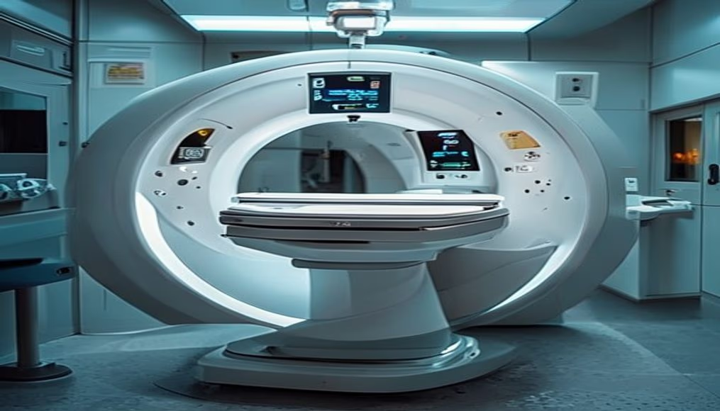
Mri scanner machine in radiology department
1344×768 · AVIF · CC BY 4.0
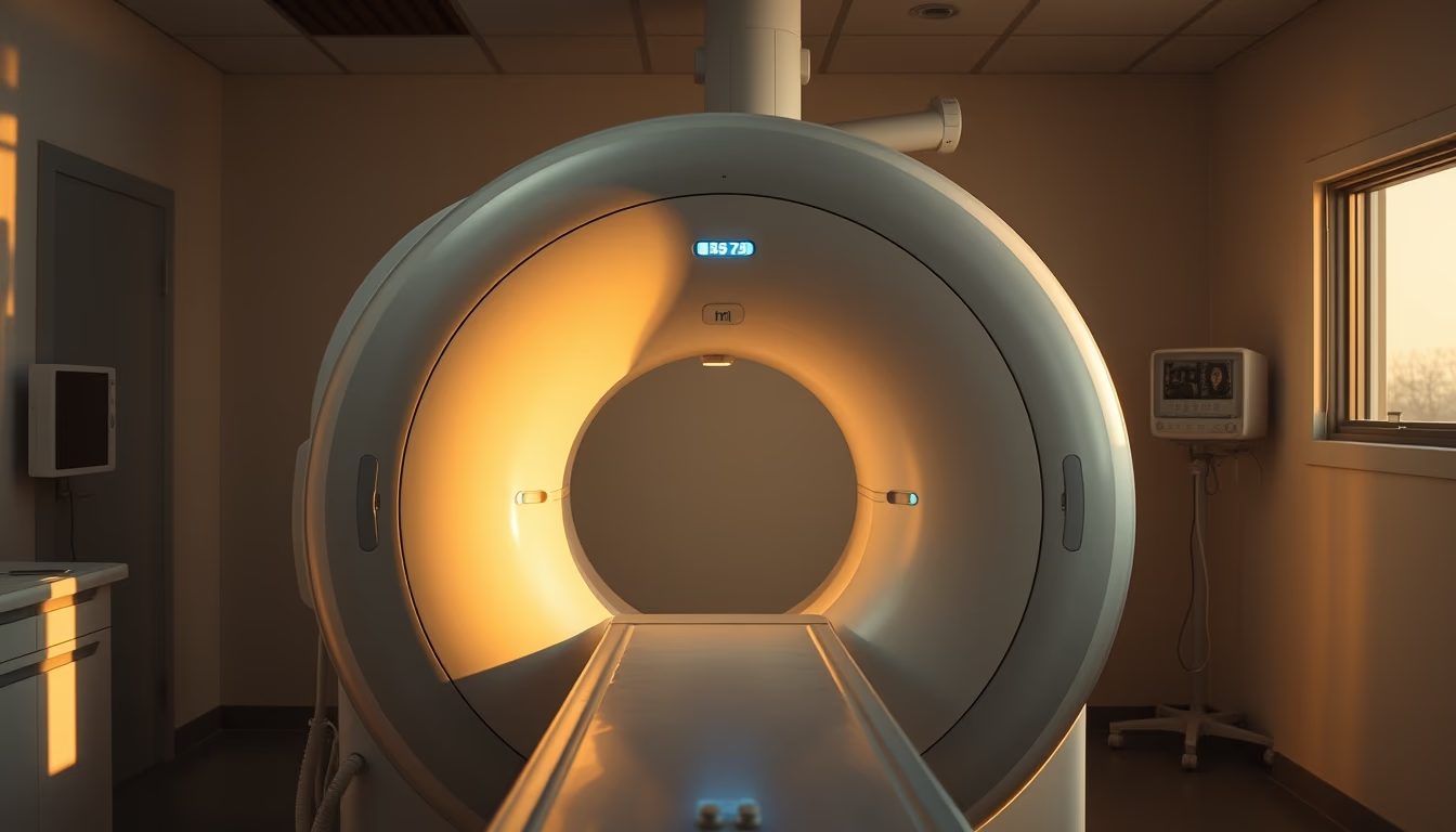
Magnetic resonance imaging is a non-invasive imaging exam that uses magnetic fields and radio waves to generate detailed images of the body's organs and tissues.
About this subject
Magnetic resonance imaging (MRI) is a diagnostic imaging technique that has revolutionized modern medicine. Unlike X-rays or computed tomography, MRI does not use ionizing radiation, making it especially safe for repeated exams and vulnerable populations such as children and pregnant women (although there are relative contraindications in the first trimester). The physical principle behind the exam is nuclear magnetic resonance: protons in the body's water molecules are aligned by an intense magnetic field (usually between 1.5 and 3 teslas, where 1 tesla is about 20,000 times the Earth's magnetic field) and then excited by radiofrequency pulses. As they return to equilibrium, they emit signals captured by antennas (coils) and processed by computers to form high-resolution three-dimensional images.
In Brazil, MRI is mainly available in large hospitals and specialized clinics, especially in the South and Southeast regions. The public health system (SUS) offers the exam, but waiting lists can be long, leading to growth in the private diagnostic imaging sector. The MRI scanner itself is a sophisticated piece of equipment that can cost between R$2 million and R$5 million, depending on magnet strength and additional features such as artificial intelligence software to speed up image acquisition. The exam is indicated for a wide range of conditions: from musculoskeletal injuries (such as ligament tears and herniated discs) to brain tumors, heart diseases, and abdominal abnormalities.
A technical curiosity is that MRI is extremely sensitive to movement: the patient must remain still for several minutes, which can be challenging for children or claustrophobic individuals. To minimize discomfort, there are open machines (with lower magnetic field) and sedation protocols. Additionally, the presence of ferromagnetic metals in the body (such as pacemakers, aneurysm clips, or metal fragments in the eyes) is an absolute contraindication, due to the risk of displacement or heating. Safety is so rigorous that before entering the exam room, the patient undergoes a detailed questionnaire and metal detection.
Historically, MRI was developed from the work of Paul Lauterbur and Peter Mansfield, who won the Nobel Prize in Physiology or Medicine in 2003. The first human exam was performed in 1977, and since then the technology has evolved rapidly: today, there are already 7-tesla machines for research, capable of generating submillimeter resolution images. In Brazil, the Brazilian Society of Radiology estimates that there are about 1,500 MRI machines in operation, concentrated mainly in São Paulo, Rio de Janeiro, and Minas Gerais.
Frequently Asked Questions
What is the difference between MRI and CT scan?
MRI uses magnetic fields and radio waves without ionizing radiation, making it ideal for soft tissues like brain and muscles. CT uses X-rays and is faster, indicated for bones and emergencies.
How long does an MRI scan take?
On average, 30 to 60 minutes, depending on the area being examined and the need for contrast. More complex exams, such as heart or abdomen, can take up to 90 minutes.
Is MRI harmful to health?
No. It is a safe and painless exam with no radiation. However, patients with metal implants, pacemakers, or claustrophobia should inform the doctor, as there may be contraindications.
Direct URL
https://pub-c7d6a6ea828543ac903a74a341ccb2e1.r2.dev/imagens/mri-scanner-machine-in-radiology-department-documentary-photography-golden-hour-late-afternoon.avifHow to credit
Include a visible link back to UtilizAí. Copy one of the snippets below:
<a href="https://xn--utiliza-eza.com/en/midia/imagens/mri-scanner-machine-in-radiology-department-documentary-photography-golden-hour-late-afternoon">Mri scanner machine in radiology department</a> by <a href="https://xn--utiliza-eza.com">UtilizAí</a>, licensed under <a href="https://creativecommons.org/licenses/by/4.0/">CC BY 4.0</a>.
[Mri scanner machine in radiology department](https://xn--utiliza-eza.com/en/midia/imagens/mri-scanner-machine-in-radiology-department-documentary-photography-golden-hour-late-afternoon) by [UtilizAí](https://xn--utiliza-eza.com), CC BY 4.0
License: CC-BY-4.0