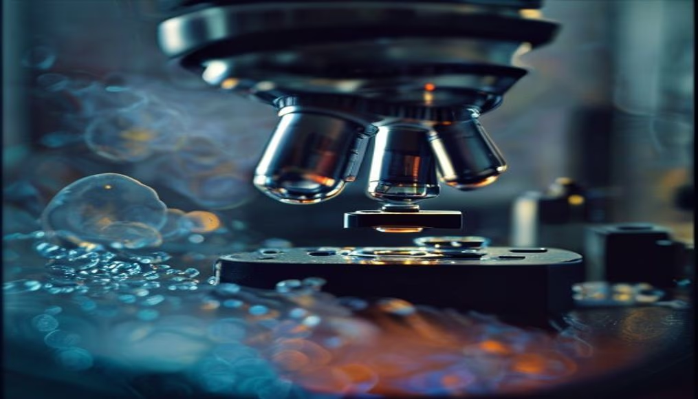
Cells under microscope
1344×768 · AVIF · CC BY 4.0
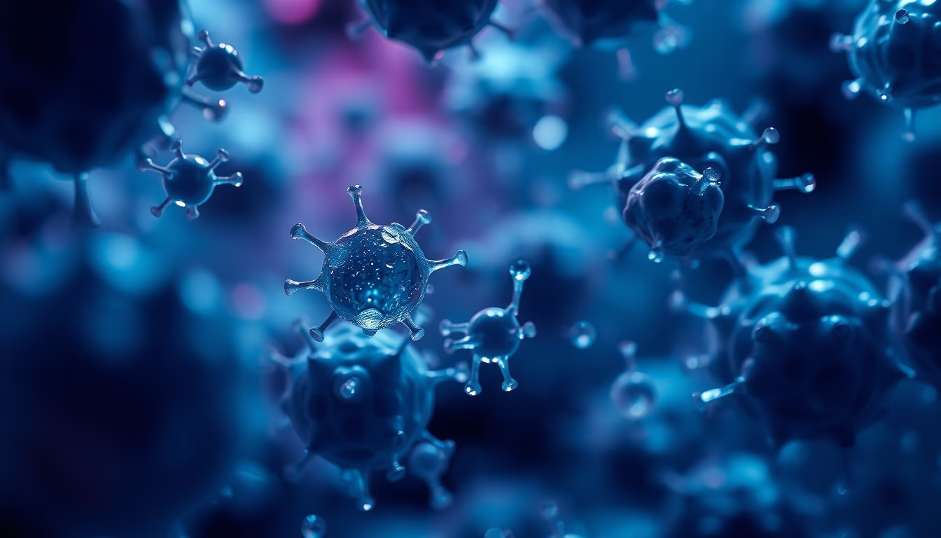
Microscopic cell images reveal the complexity of life on a small scale.
About this subject
Cells are the basic units of life, and observing them under a microscope is a fascinating experience. The image shows cells in different stages of division, with internal structures such as nucleus, mitochondria, and ribosomes. Optical and electron microscopy have enabled major advances in understanding cell biology. Historically, Antonie van Leeuwenhoek is considered the father of microscopy, having discovered microorganisms and cells in 1676.
Frequently Asked Questions
What can be seen in a microscope image of cells?
Cell structures such as nucleus, mitochondria, ribosomes, and cell membranes can be seen.
Who was the first to observe cells under a microscope?
Antonie van Leeuwenhoek is considered the father of microscopy and discovered microorganisms and cells in 1676.
What are the main microscopy techniques used to observe cells?
The main techniques are optical microscopy and electron microscopy, which allow cells to be observed at different scales and resolutions.
Direct URL
https://pub-c7d6a6ea828543ac903a74a341ccb2e1.r2.dev/imagens/cells-under-microscope-fine-art-photography-v7.avifHow to credit
Include a visible link back to UtilizAí. Copy one of the snippets below:
<a href="https://xn--utiliza-eza.com/en/midia/imagens/cells-under-microscope-fine-art-photography-v7">Cells under microscope</a> by <a href="https://xn--utiliza-eza.com">UtilizAí</a>, licensed under <a href="https://creativecommons.org/licenses/by/4.0/">CC BY 4.0</a>.
[Cells under microscope](https://xn--utiliza-eza.com/en/midia/imagens/cells-under-microscope-fine-art-photography-v7) by [UtilizAí](https://xn--utiliza-eza.com), CC BY 4.0
License: CC-BY-4.0